POPULARITY

3 episodes with efast

2 episodes with efast

2 episodes with efast

2 episodes with efast

2 episodes with efast

2 episodes with efast

CoROM cast. Wilderness, Austere, Remote and Resource-limited Medicine.
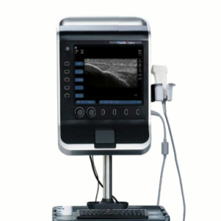
This week, Aebhric O'Kelly speaks with three combat medics from Tactical Medicine North following a Tactical APUS instructor development programme in Malta. The discussion explores whether ultrasound can be taught to non-medical personnel operating in combat environments, including Combat Lifesavers (CLS) and Combat Medic Corpsmen (CMC), and how ultrasound may support prolonged casualty care, triage, and telemedicine in Ukraine. The conversation challenges traditional assumptions regarding ultrasound education, introduces the Tactical APUS concept, discusses modifications to the standard eFAST examination sequence, and reviews preliminary observations from a study comparing parasternal long-axis (PLAX) and subxiphoid cardiac views. Chapters00:00 – Introduction01:06 – Can Non-Medics Learn Ultrasound?03:00 – Lessons from the APUS Course05:30 – The Power of Home Points07:50 – What is Tactical APUS?10:00 – Adapting eFAST for Combat Operations12:30 – Hypothermia Prevention During Ultrasound15:20 – The Controversial Change: Heart Last20:00 – PLAX vs Subxiphoid Cardiac Views24:40 – Teaching Maltese Nurses29:10 – Should We Teach Ultrasound to Combat Lifesavers?32:20 – Ultrasound as a Triage Tool35:10 – Advice for Future Tactical Ultrasound Providers38:00 – Closing RemarksKey TakeawaysThe parasternal long-axis cardiac viewappears easier for novice learners than the traditional subxiphoid view.Overview of the APUS and Tactical APUS training programme conducted in Malta. Discussion on teaching eFAST ultrasound to Combat Lifesavers and Combat Medic Corpsmen.Comparison with early challenges teaching combat medicine to personnel without formal medical backgrounds. Importance of simple teaching techniques and instructor adaptability.Introduction of the "Home Point" concept for each eFAST window. How home points help students recover when they become disoriented during scanning.Development of a one-day ultrasoundcurriculum for tactical providers.Focus on eFAST as a trauma tool for prolonged field care and telemedicine support.Discussion of modifying the traditional eFAST sequence.Prioritising lung assessment over cardiac views.The dangers of exposing casualties during scanning.Importance of maintaining casualty insulation and minimising gel exposure.Why the Tactical APUS team moved cardiac assessment after lung assessment.Students consistently finding the parasternal long-axis view easier to obtain.Experience using Maltese nurses as pilot students.Differences between teaching healthcare professionals and non-medical personnel.Language barriers and instructional adaptations. Moving beyond "Can we?" to "Should we?"Ultrasound as a prolonged casualty care and telemedicine tool.Supporting decision-making during extended evacuations. Using eFAST to prioritise casualties during mass casualty situations.Early identification of internal bleedingand pneumothorax.Potential role of optic nerve sheath diameter (ONSD) assessment in blast-related head injuries. Importance of accessibility of handheld ultrasound devices.The role of deliberate practice and repetition in ultrasound mastery. Reflections on the success of the Tactical APUS pilot programme.Future collaboration between CoROM and Tactical Medicine North.Final thoughts from the Ukrainian instructors. Ultrasound can be successfully taught to Combat Lifesavers and Combat Medic Corpsmen when training is focused on pattern recognition and image acquisition rather than advanced interpretation."Home Points" provide a powerful cognitive aid for novice sonographers.Lung ultrasound may provide greater battlefield utility than cardiac ultrasound because interventions can be performed immediately.Hypothermia prevention must remainintegrated into all ultrasound training and operational use.

Karl Alvarsson er fyrrverandi flugumferðarstjóri og lögfræðingur og hefur unnið ýmis sérfræðistörf tengd fluginu. Hann er í dag formaður Fagráðs um flugmál sem er innviðaráðherra til ráðuneytis í málaflokknum og er þar að auki formaður skipulagsnefndar fyrir Keflavíkurflugvöll. Í þættinum er rætt við Karl um skipulagsmálin, þá einkum í Reykjavík. Karl gagnrýnir að alþjóðaflugvöllurinn í höfuðborginni, sem jafnrframt er hjartað í innanlandssamgöngukerfinu, skuli háður starfsleyfi frá heilbrigðiseftirliti borgarinnar. Hann ræðir líka um fyrirkomulag skipulagsmála á Keflavíkurflugvelli og um helstu verkefni sem þar eru framundan. Ásælni flugfélaga í að hafa leyfi til flugrekstrar á Möltu eru líka til umræðu í þættinum og þeirri spurningu varpað fram hvort íslensk stjórnvöld geti gert eitthvað til að efla íslenska flugrekendur í alþjóðlegri samkeppni. Áhugavert spjall fyrir alla þá sem vilja fylgjast með í flugmálum.

Send us a textExploring Plant-Based Nutrition and Implementing an eFastIn this episode, Michelle and Dan discuss their experience with Michael Greger's plant-based cookbooks, "How Not to Die" and "How Not to Diet." They highlight the importance of whole food plant-based meals for nutritional health and managing cholesterol. The conversation then shifts to the concept of an eFast, which involves intentionally taking breaks from electronic devices to improve sleep and overall well-being. They explore the challenges of compulsive and impulsive behaviors associated with device use, and provide practical tips for successfully implementing an eFast. Lastly, they discuss the interconnectedness of diet, sleep, and technology use.00:00 Introduction and Technical Difficulties00:19 Reviewing Michael Greger's Cookbooks01:50 Transition to Whole Food Plant-Based Diet02:02 Challenges of Grocery Shopping for Whole Foods05:00 Benefits of Plant-Based Diet for Sleep06:23 Introduction to eFasting09:49 Understanding Compulsive and Impulsive Behaviors14:30 Implementing eFasting for Better Sleep17:27 Conclusion and Final ThoughtsTheme music "Happy Days by FSM Team" courtesy of https://www.free-stock-music.com Support the showPlease go to the following page to support the show: https://www.buzzsprout.com/1692604/support www.youtube.com/@sleeptakeoutwww.danielbaughn.comwww.dosleep.comsleeptakeout@gmail.com

Time to enter the Echo Chamber! Dr Mai Nguyen joins Callum to discuss eFAST in Trauma in this bonus episode. Following on from a very detailed discussion of all things trauma in lasts week's main episode, we're diving into the specifics of using POCUS in trauma, and importantly how to do it effectively and efficiently. Dr Nguyen is a Consultant in Emergency Medicine at University Hospital Limerick where she is the Director of Ultrasound Training for Emergency Medicine, an EGLS instructor and a Level 1 Ultrasound Trainer. In short, the perfect guest to run through all things eFAST. If you didn't want to build your POCUS skills before, you sure will after this episode! Listen along with the show notes and get involved in the conversation on Twitter and Instagram.

In this episode we continue our Pediatric Emergency Medicine Assembly(PEM23) series with the evidence and growth of contract enhanced ultrasound in the emergency department. In this episode, we talk about the FAST and eFAST exams as well as some of the "how does it work" of contrasted imaging. Use the code EMPOWER to save $100 on registration for ACEP's Leadership & Advocacy Conference - Register Now LAC...https://www.acep.org/lac/

Dominos

TestTalks | Automation Awesomeness | Helping YOU Succeed with Test Automation
Is there still value in keyword-driven approaches to automation? In this episode, Marcus Thomas, founder of Quart Consulting, shares his experience helping hundreds of folks with their automation testing frameworks. Discover why we loved WinRunner, how to leverage time-tested techniques for automation, why a developer-heavy approach to automation is not always the best way, and how to use eFAST to create and execute automated tests quickly.

It's the JournalFeed Podcast for the week of March 7-11, 2022. We cover eFAST for pneumothorax, advanced airway vs bag-valve mask for OHCA, muscle relaxants for back pain, ongoing opioid use after fentanyl in the ED, and heart rate and mortality risk in PE.
In this podcast we discuss the indications of x-rays, eFAST, CT scans, contrast studies and other investigations. Attached to the podcast is a PDF with links to additional resources. Stanleur Capital: Medical practice and personal financial solutions · Additional material Radiology in the trauma resuscitation room.pdf — PDF (31.1 KB)
In this podcast we discuss the indications of x-rays, eFAST, CT scans, contrast studies and other investigations. Attached to the podcast is a PDF with links to additional resources. Additional material Radiology in the trauma resuscitation room.pdf — PDF (31.1 KB)

Trauma Resuscitation and the Covid-19 Pandemic in South Africa In this podcast, Roger Harris interviews Victoria Stephen about her experience as an emergency physician in a regional South African hospital. Sadly, trauma resuscitation is a big part of working in Emergency Medicine in South Africa. Blunt force assaults and stab wounds are regular presentations. However, July 2021 was unlike anything Doctor Victoria Stephen had ever experienced. In July, South Africa was deep into its' third wave of Covid-19 infections. Vaccination rates were low and there was a huge burden of Covid patients in the Emergency Department. The ICU was completely overwhelmed, making this by far the worst of the pandemic that they had seen to date. To compound this, piped oxygen levels were running desperately low. The hospital relied on daily oxygen deliveries to keep Covid patients alive. Moreover, to add to the challenge, political unrest broke out and quickly escalated to riots with extreme violence across South Africa. At the time the violence erupted, Tori had over 120 Covid patients in the hospital. Added to this the Trauma resuscitation was managing approximately 34 patients with gunshot wounds per day. With just four doctors working at night and six doctors working during the day, Tori's team scrambled to manage an overwhelming number of high acuity patients. For the first time in her career, Tori found herself frightened for her safety. Having grown up in South Africa, Tori was no stranger to avoiding danger but this felt very out of control. The thought of managing a busy emergency department inundated with trauma patients in the middle of the covid pandemic is frightening enough for most of us, but to do so in such a resource-limited environment with so few nurses and doctors is truly incredible. Tori believes Emergency Medicine training in South Africa prepares the team to function under such pressure. She believes that the team knows that the lack of resources means they must all pull together. Their training is diverse enough that they have the mental and clinical skills to step up and of course as an ultrasound geek Tori adds that EFAST scanning has a big role to play. Tori is a humble but inspirational clinician on the frontline of providing care in a volatile environment and she believes we can all learn something from her experience. Tune in to a compelling conversation with one of our favourites. Trauma Resuscitation and the Covid-19 Pandemic in South Africa Finally, for more like this head to our podcast page #CodaPodcast

In this session, we interview Gaynor Prince. Gaynor is an emergency physician based in New Zealand and has developed a subspecialty interest in ultrasound in ED. We take a look at point of care ultrasound, its utility and its limitations. We will be especially focussing on the EFAST and how it has been adaptive and progressive in point of care treatment in the past 5 years. We also take a look at ultrasound probes and the positive and negative interaction with tissue. Gaynor unpacks some of the fundamental advantages and disadvantages of POCUS and how it has been adapted, been made portable and democratised amongst clinicians in recent years. We unpack the principles of EFAST, what we are looking for, when to look and where. We examine the difference image representations of fluid, blood, ascites, urine, intestinal contents, lungs, air. Gaynor them looks at the anatomical variations and how to optimise the view, interplay with clinical questioning, repetition of scans and preferential windows to see the anatomy. We go sequentially through the EFAST and look at tips and tricks from Gaynor's practice and how these can be related to everyone's ultrasound practice and decision making. We finally look at a prime example of when USS has been really useful to Gaynor's practice in one of the remotest and most extreme environments - Antarctica. She recalls a story of when ultrasound greatly assisted her decision making, illustrating some of the unique examples of this modality in remote and austere locations. You can see more from Gaynor here: https://www.wem.academy/videos/extreme-medicine-research/ultrasoundinextremeenvironments/ This episode has been kingly re-shared from WEMcast. World Extreme Medicine provide courses, resources, training and conferences and can be found at: https://worldextrememedicine.com Please enjoy the episode.

In this session, we interview Gaynor Prince. Gaynor is an emergency physician based in New Zealand and has developed a subspecialty interest in ultrasound in ED. We take a look at point of care ultrasound, its utility and its limitations. We will be especially focussing on the EFAST and how it has been adaptive and progressive in point of care treatment in the past 5 years. We also take a look at ultrasound probes and the positive and negative interaction with tissue. Gaynor unpacks some of the fundamental advantages and disadvantages of POCUS and how it has been adapted, been made portable and democratised amongst clinicians in recent years. We unpack the principles of EFAST, what we are looking for, when to look and where. We examine the difference image representations of fluid, blood, ascites, urine, intestinal contents, lungs, air. Gaynor them looks at the anatomical variations and how to optimise the view, interplay with clinical questioning, repetition of scans and preferential windows to see the anatomy. We go sequentially through the EFAST and look at tips and tricks from Gaynor's practice and how these can be related to everyone's ultrasound practice and decision making. We finally look at a prime example of when USS has been really useful to Gaynor's practice in one of the remotest and most extreme environments - Antarctica. She recalls a story of when ultrasound greatly assisted her decision making, illustrating some of the unique examples of this modality in remote and austere locations. You can see more from Gaynor here: https://www.wem.academy/videos/extreme-medicine-research/ultrasoundinextremeenvironments/

Core Questions What are the injuries for the following blunt trauma mechanisms: Head-on collision Rear end collision Lateral (T-bone) collision Rollover Ejected from vehicle Windshield damage Steering wheel damage Dashboard involvement or damage Restraint or seat belt use Air bag deployment Low-speed pedestrian versus automobile High-speed pedestrian versus automobile Bicycle versus automobile Non-automobile-related Vertical impact falls Horizontal impact falls Outline an approach to the primary survey for the trauma patient. Describe the elements of the eFAST exam. Outline an approach to the secondary survey in the trauma patient. Detail relevant ancillary laboratory tests to order in the trauma patient. Canadian CT Head Rule Canadian C-Spine Rule NEXUS C-Spine Rule NEXUS Chest Rule List the components of the following imaging decision-making tool What are the indications for a CT abdomen/pelvis in the trauma patient? Wisecracks What are the mechanisms of injury for the following weapons: Knives Handgun rounds Shotgun rounds Rifle rounds What is the LD50 in feet for falls from a given height? What is permissive hypotension and what evidence does it have?

Core Questions What are the injuries for the following blunt trauma mechanisms: Head-on collision Rear end collision Lateral (T-bone) collision Rollover Ejected from vehicle Windshield damage Steering wheel damage Dashboard involvement or damage Restraint or seat belt use Air bag deployment Low-speed pedestrian versus automobile High-speed pedestrian versus automobile Bicycle versus automobile Non-automobile-related Vertical impact falls Horizontal impact falls Outline an approach to the primary survey for the trauma patient. Describe the elements of the eFAST exam. Outline an approach to the secondary survey in the trauma patient. Detail relevant ancillary laboratory tests to order in the trauma patient. Canadian CT Head Rule Canadian C-Spine Rule NEXUS C-Spine Rule NEXUS Chest Rule List the components of the following imaging decision-making tool What are the indications for a CT abdomen/pelvis in the trauma patient? Wisecracks What are the mechanisms of injury for the following weapons: Knives Handgun rounds Shotgun rounds Rifle rounds What is the LD50 in feet for falls from a given height? What is permissive hypotension and what evidence does it have?
Váá hvað gaslighting er lúmsk og ömurleg hegðun. Þetta er umræðuefni sem kom virkilega á óvart og við höldum að margir geti lært helling af. Skoðum!
Váá hvað gaslighting er lúmsk og ömurleg hegðun. Þetta er umræðuefni sem kom virkilega á óvart og við höldum að margir geti lært helling af. Skoðum!
Come for a hands-on session with ultrasound! We will be going over the basic principles, application via the eFAST exam (Focused Assessment with Ultrasound in Trauma), and hands on time with procedural models. We will also be providing IV, LP, and Central Line Models to practice on. All are welcome. This is a basic introduction to ultrasound.
Contributor: Aaron Lessen, MD Educational Pearls:. Focused assessment with Sonography for Trauma (FAST) exam and the extended-FAST (eFAST) are essential components of current trauma care and evaluation There has been an accumulation of research to provide an estimate of effectiveness of identifying certain injuries with ultrasound: For identifying a pneumothorax, the sensitivity ~70% and specificity ~99%. For pericardial effusions, sensitivity 90% and specificity ~ 94%. For hemoperitoneum, sensitivity ~74% and the specificity ~98%. While ultrasound is excellent for identifying many injuries, it may not be adequate alone to rule out serious injuries if the clinical suspicion is high based on these pooled studies References Netherton S, Milenkovic V, Taylor M, Davis PJ. Diagnostic accuracy of eFAST in the trauma patient: a systematic review and meta-analysis. CJEM. 2019;21(6):727‐738. doi:10.1017/cem.2019.381 Summarized by Jackson Roos, MS4 | Edited by Erik Verzemnieks, MD
Contributor: Aaron Lessen, MD Educational Pearls:. Focused assessment with Sonography for Trauma (FAST) exam and the extended-FAST (eFAST) are essential components of current trauma care and evaluation There has been an accumulation of research to provide an estimate of effectiveness of identifying certain injuries with ultrasound: For identifying a pneumothorax, the sensitivity ~70% and specificity ~99%. For pericardial effusions, sensitivity 90% and specificity ~ 94%. For hemoperitoneum, sensitivity ~74% and the specificity ~98%. While ultrasound is excellent for identifying many injuries, it may not be adequate alone to rule out serious injuries if the clinical suspicion is high based on these pooled studies References Netherton S, Milenkovic V, Taylor M, Davis PJ. Diagnostic accuracy of eFAST in the trauma patient: a systematic review and meta-analysis. CJEM. 2019;21(6):727‐738. doi:10.1017/cem.2019.381 Summarized by Jackson Roos, MS4 | Edited by Erik Verzemnieks, MD

Það má efast..ekki efast um það by Rögnvaldur Hreiðarsson
Come for a hands-on session with ultrasound! We will be going over the basic principles, application via the eFAST exam (Focused Assessment with Ultrasound in Trauma), and hands on time with procedural models. We will also be providing IV, LP, and Central Line Models to practice on. All are welcome. This is a basic introduction to ultrasound.

Sigurbjörn Daði Dagbjartson settist niður með Jóhanni Árna Ólafssyni þjálfara Grindavíkur og ræddi við hana um komandi tímabil í Dominos deild kvenna.Er upptakan í röð þar sem að sest er niður með þjálfurum og spáð í spilin fyrir komandi tímabilUpptakan er hluti af Sibbaspjalli, en það er bæði aðgengilegt sem podcast inni á iTunes og öðrum veitum, sem og á myndbandi hér fyrir neðan.

Most of us struggle with overuse of electronic devices. They are so convenient and address so many issues all in one place. But most of us also know that such things can become addictive and keep us locked in to them and away from the walk God has planned for us. But we can't just put the cell phone in the drawer for a month. We need it everyday to interact with others, conduct business, or find needful things. However, if we can learn the value of daily intermittent fasting, creating ever increasing pockets of hours away from the device, we will instantly see more meaningful things fill that space and regain control of our time and lives!- How to assess if you spend too much time on your cell phone or ipad- Why a 30 Day Fast is beneficial but also may not be reasonable for everyone.- How the Intermittent eFast approach can be the daily answer for you.- What are the immediate benefits to ever increasing hours without electronics- How to walk through a daily with intervals of focus and peace and without the phone

Show Notes Jeff: Welcome back to EMplify, the podcast corollary to EB Medicine’s Emergency Medicine Practice. I’m Jeff Nusbaum, and I’m back with my co-host, Nachi Gupta. This month, after a few months of primarily medical topics, we’re talking trauma, specifically Blunt Cardiac Injury: Emergency Department Diagnosis and Management. Nachi: With no gold standard diagnostic test and with complications ranging from simple ectopic beats to fulminant cardiac failure and death, this isn’t an episode you’ll want to miss. Jeff: Before we begin, let me give a quick shout out to our incredible group of authors from New York -- Dr. Eric Morley, Dr. Bryan English, and Dr. David Cohen of Stony Brook Medicine and Dr. William Paolo, residency program director at SUNY Upstate. I should also mention their peer reviewers Drs. Jennifer Maccagnano and Ashley Norse of the NY institute of technology college of osteopathic medicine and UF Health Jacksonville, respectively. Nachi: This month’s team parsed through roughly 1200 articles as well as guidelines from the eastern association for surgery in trauma also known as EAST. Jeff: Clearly a large undertaking for a difficult topic to come up with solid evidence based recommendations. Nachi: For sure. Let’s begin with some epidemiology, which is admittedly quite difficult without universally accepted diagnostic criteria. Jeff: As you likely know, despite advances in motor vehicle safety, trauma remains a leading cause of death for young adults. In the US alone, each year, there are about 900,000 cases of cardiac injury secondary to trauma. Most of these occur in the setting of vehicular trauma. Nachi: And keep in mind, that those injuries don’t occur in isolation as 70-80% of patients with blunt cardiac injury sustain other injuries. This idea of concomitant trauma will be a major theme in today’s episode. Jeff: It certainly will. But before we get there, we have some more definitions to review - cardiac concussion and contusion, both of which were defined in a 1989 study. In this study, cardiac concussion was defined as an elevated CKMB with a normal echo, while a cardiac contusion was defined as an elevated CKMB and abnormal echo. Nachi: Much to my surprise, though, abnormal echo and elevated ck-mb have not been shown to be predictive of adverse outcomes, but conduction abnormalities on ekgs have been predictive of development of serious dysrhythmia Jeff: More on complications in a bit, but first, returning to the idea of concomitant injuries, in one autopsy study of nearly 1600 patients with blunt trauma - cardiac injuries were reported in 11.9% of cases and contributed to the death of 45.2% of those patients. Nachi: Looking more broadly at the data, according to one retrospective review, blunt cardiac injury may carry a mortality of up to 44%. Jeff: That’s scary high, though I guess not terribly surprising, given that we are discussing heart injuries due to major trauma... Nachi: The force may be direct or indirect, involve rapid deceleration, be bidirectional, compressive, concussive, or even involve a combination of these. In general, the right ventricle is the most frequently injured area due to the proximity to the chest wall. Jeff: Perfect, so that's enough background, let’s talk differential. As you likely expected, the differential is broad and includes cardiovascular injuries, pulmonary injuries, and other mediastinal injuries like pneumomediastinum and esophageal injuries. Nachi: Among the most devastating injuries on the differential is cardiac wall rupture, which not surprisingly has an extremely high mortality rate. In terms of location of rupture, both ventricles are far more likely to rupture than the atria with the right atria being more likely to rupture than the left atria. Atrial ruptures are more survivable, whereas complete free wall rupture is nearly universally fatal. Jeff: Septal injuries are also on the ddx. Septal injuries occur immediately, either from direct impact or when the heart becomes compressed between the sternum and the spine. Delayed rupture can occur secondary to an inflammatory reaction. This is more likely in patients with a prior healed or repaired septal defects. Nachi: Valvular injuries, like septal injuries, are rare. Left sided valvular damage is more common and carries a higher mortality risk. In order, the aortic valve is more commonly injured followed by the mitral valve then tricuspid valve, and finally the pulmonic valve. Remember that valvular damage can be due to papillary muscle rupture or damage to the chordae tendineae. Consider valvular injury in any patient who appears to be in cardiogenic shock, has hypotension without obvious hemorrhage, or has pulmonary edema. Jeff: Next on the ddx are coronary artery injuries, which include lacerations, dissections, aneurysms, thrombosis, and even MI secondary to increased sympathetic activity and platelet activity after trauma. In one review, dissection was the most commonly uncovered pathology, occurring 71% of the time, followed by thrombosis, which occured only 7% of the time. The LAD is the most commonly injured artery followed by the RCA. Nachi: Pericardial injury, including pericarditis, effusion, tamponade, and rarely rupture, is also certainly on the differential. Jeff: In terms of dysrhythmias, sinus tachycardia is the most common dysrhythmia, with other rhythms, including PVC / PAC / and afib being found only 1-6% of the time. Nachi: And while conduction blocks are rare, a RBBB is the most commonly noted, followed by a 1st degree AVB. Jeff: Though also rare, commotio cordis deserves it’s own section as its the second most common cause of death in athletes < 18 who are victims of blunt trauma. Though only studied in swine models, it’s hypothesized that the impact to the chest wall during T-wave upstroke can precipitate v-fib. Nachi: Aortic root injuries usually occur at the insertion of the ligamentum arteriosum and isthmus. Such injuries typically result in aortic insufficiency. Jeff: And the last pathology on the differential requiring special attention is a myocardial contusion. Again, no standard definition exists, with some diagnostic criteria including simply chest pain and increasing cardiac enzymes, and others including cardiac dysfunction, ecg abnormalities, wall motion abnormalities, and an elevation of cardiac enzymes. Nachi: Certainly a pretty broad differential… before moving on to the work up, Jeff why don’t you get us started with prehospital care? Jeff: Prehospital management should focus on rapid identification and stabilization of life threatening injuries with expeditious transport as longer prehospital times have been associated with increased mortality in trauma. Immediate transport to a Level I trauma center should be the highest priority for those with suspected blunt cardiac injury. Nachi: In terms of who specifically should be transporting the patient, a Cochrane review evaluated the utility of ALS vs BLS transport in trauma. There is reasonably good data to support BLS over ALS, even when controlling for trauma severity. Moreover, when airway management is needed, advanced airway techniques by ALS crews were associated with decreased odds of survival. Regardless of who is there, the message is the same: focus not on interventions, but instead on rapid transport. Jeff: And if it does happen to be an ALS transport crew, without delaying transport, pain management with fentanyl is both safe and reasonable and preferred over morphine. Post opiate hypotension in prehospital trauma patients is a rare but documented complication. Nachi: And if the prehospital team is lucky enough, or maybe unlucky enough, i don’t know, to have a credentialed provider who can perform ultrasound for those suspected of having a blunt cardiac injury, the general prehospital data on ultrasound is sparse. As of now, it’s difficult to conclude if prehospital US improves care for trauma patients. Jeff: Interestingly, the system I work in has prehospital physicians, who do carry US, but I can’t think of a major trauma where ultrasound changed any of the decisions we made. Nachi: Right, and I think that just reinforces the main point here: there may be a role, we just don’t have the data to support it at this time. Jeff: Great, let’s move onto ED care, beginning with the H&P. Nachi: On history, make sure to elucidate if there is any chest pain, and if it’s onset was before or after the traumatic event. In addition, make sure to ask about dyspnea, fatigue, palpitations, and lightheadedness. Jeff: And don’t forget to get the crash details from the EMS crew before they depart! As a side note, for anyone taking oral boards in a few months, don’t forget to ask the EMS crew for the details!!! Nachi: A definite must for oral boards and for your clinical practice. Jeff: In terms of the physical, tachycardia is the most common abnormality in blunt cardiac injury. In those with severe injury, you may note refractory hypotension secondary to cardiogenic shock. But don’t be reassured by normal vitals, especially in the young, who may be compensating well despite being quite ill. Nachi: Fully undress the patient to appropriately inspect and percuss the chest wall - looking for signs of previous cardiac surgeries or pacemaker placement, as well as to auscultate for new murmurs which may be a sign of valvular injury. Jeff: Similarly, as concomitant injuries are common, inspect the abdomen, looking for ecchymosis patterns, which often accompany blunt cardiac injury. Nachi: Pretty standard stuff. Let’s move on to diagnostic testing. Jeff: Lab testing should include a CBC, BMP, coags, troponin, lactate, and T&S. In one retrospective analysis, an elevated troponin and a lactate over 2.5 were predictors of mortality. Nachi: Additionally, in patients with chest trauma, a troponin > 1.05 was associated with a greater risk for dysrhythmias and LV dysfunction. Jeff: And it likely goes without saying, but an EKG is a must on all trauma patients with suspicion for blunt cardiac injury in accordance with the EAST guidelines. New EKG findings requires admission for monitoring. Unfortunately, on the flip side, an ECG cannot be used to rule out blunt cardiac injury. Nachi: Diving a bit deeper into the data, in a prospective study of 333 patients with blunt thoracic trauma, serial EKG and troponins at 0, 4, and 8 hours post injury had a sensitivity and specificity of 100% and 71%, respectively. However, of those with abnormal findings, all but one had them on initial testing, leading to a negative predictive value of 98%. Jeff: Well that’s an impressive NPV and has huge implications, especially in the era of heavily monitored lengths of stay... Nachi: Definitely. In terms of radiography, a chest x-ray should be obtained as rib fractures, hemopneumothorax, and mediastinal free air are all things you wouldn't want to miss and are also associated with blunt cardiac injury. Jeff: Keep in mind, however, that the chest x-ray should not be seen as a test for pericardial fluid as up to 200 mL of fluid can be contained in the pericardial space and remain undetectable by chest radiograph. Nachi: Which is why you’ll have to turn to our good friend the ultrasound, for more useful data. The data is strong that in the hands of trained Emergency Clinicians, when parasternal, apical, and subcostal views are obtained, US has an accuracy of 97.5% for pericardial effusion. Jeff: Not only is US accurate, it’s also quick. In one RCT, the FAST exam reduced the time from arrival in the ED to operative care by 64% in the setting of trauma. Nachi: That’s impressive -- for expediting patient care and for managing ED flow. Jeff: Exactly. The authors do note however that hemopericardium is a rare finding, so, while not the focus of this article, the real utility of the FAST exam may be in its expanded form, the eFAST, in which a rapid bedside ultrasonographic lung exam for pneumothorax is included, as this can lead to immediate changes in management. Nachi: And assuming you do your FAST or eFAST and have no management changing findings, CT will often be your next test. Jeff: Yeah, EKG-gated multidetector CT can easily diagnose myocardial rupture, pneumopericardium, pericardial rupture, hemopericardium, coronary artery insult, ventricular septal defects and even valvular dysfunction. Unfortunately, CT does not perform well for the evaluation of myocardial contusions. Nachi: This is all well and good, and certainly accurate, but let’s not forget that hemodynamically unstable trauma patients, like those with myocardial rupture, need to be in the operating room, not the CT scanner. Jeff: An important point that should not be understated. Nachi: And the last major testing modality to discuss is the echocardiogram. Jeff: The echo is a fantastic test for detecting focal cardiac dysfunction often see with cardiac contusions, hemopericardium, and valve disruption. Nachi: And it’s worth noting that transthoracic is enough, as transesophageal, despite the better images, hasn’t been shown to change management. TEE should be saved for those in whom a optimal TTE study isn’t feasible. Jeff: Great point. And one last quick note on echo: in terms of guidelines, the EAST guidelines from 2012 specifically recommend an echo in hemodynamically unstable patients or those with a persistent new dysrhythmia without other sources of ongoing hemorrhage or neurologic etiology of instability. Nachi: Perfect, so that wraps up testing and imaging for our blunt cardiac injury patient. Let’s move on to treatment. Jeff: In terms of initial resuscitation, there is an ever increasing body of literature to support blood transfusion over crystalloid in patients requiring volume expansion in trauma. There are no specific guidelines for transfusion in the setting of blunt cardiac injury, so stick to your standard trauma protocols. Nachi: It is worth noting, though, that there is literature outside of trauma for those with pericardial effusions, suggesting that those with a SBP < 100 have substantial benefit from volume expansion. So keep this in mind if your clinical suspicion is high and your trauma patient has a soft but not truly shocky blood pressure. Jeff: Operative management, specifically ED thoracotomy is a heavily debated topic, and it’s next on our list to discuss. Nachi: The 2015 EAST guidelines conditionally recommend ED thoracotomy for moribund patients with signs of life. The Western Trauma Association broadens the ED thoracotomy window a bit to include anyone with no signs of life but less than 10 minutes of CPR. The latter also recommend ED thoracotomy in those with refractory shock. Jeff: Though few studies exist on the topic, in one study of 187 patients, cardiac motion on US was 100% sensitive for predicting survivors. Nachi: Not great data, but it does support one's decision to stop any further work up should there be no cardiac activity, which is important, because the decision to pursue an ED thoracotomy is not an easy one. Jeff: And lastly, emergent pericardiocentesis may be another option in an unstable patient when definitive operative management is not possible. But do note that pericardiocentesis is only a temporizing measure, and not definitive for cardiac tamponade. Nachi: Treatment for dysrhythmias is standard, treat in accordance with standard ACLS protocols, as formal randomized trials on prophylaxis and treatment in the setting of blunt cardiac injury do not exist. Jeff: Seems reasonable enough. And in the very rare setting of an MI after blunt cardiac injury, you should involve cardiology, cardiothoracic surgery, and trauma to help make important management decisions. Data is, again, lacking, but the patient likely needs percutaneous angiography for appropriate diagnosis and potentially further intervention. Definitely hold off on ASA and likely nitroglycerin, at least until significant bleeding has been ruled out. Nachi: Yup, no style points for giving aspirin to a bleeding trauma patient. Speaking of medications, the last treatment modality to discuss here is pain control. Pain management is essential with chest injuries, as appropriate pain management has been shown to reduce mortality in pulmonary related complications. Jeff: And in line with every acute pain consult note I’ve ever come across, a multimodal approach utilizing opioids and nonopioids is recommended. Nachi: Perfect, so that sums up treatment, next we have one special circumstance to discuss: sternal fractures. Cardiac contusions are found in 1.8-2.4% of patients with sternal fractures, almost all of which were seen on CT and not XR according to the NEXUS chest CT study. Of these patients, only 2 deaths occured, both due to cardiac causes. Thus, in patients with isolated sternal fractures, negative trops, ekg, and negative cxr - the patient can likely be discharged from the ED, as long as their pain is well-controlled. Jeff: And let’s talk controversies for this issue. We only have one to discuss: MRI. Nachi: The fact that MRI produces awesome images is not controversial, see figure 3. It’s role, however, is. In accordance with EAST guidelines, MRI may be most useful in differentiating acute ischemia from blunt cardiac injury in those with abnormal ECGs, elevated enzymes, or abnormal echos. It’s use in the hyperacute evaluation, however, is limited, in large part owing to the length of time required to complete an MRI Jeff: What a time to be alive that we even have to say that MRIs may not have a hyperacute role in trauma - absolutely crazy... Nachi: Moving on to disposition: any patient with aortic, pericardial, or myocardial injury and hemodynamic instability needs operative evaluation and likely intervention, so do not hesitate to get the consults coming or the helicopter in the air should such a patient arrive at your non-trauma center. Jeff: And in those that are hemodynamically stable, with either a positive ECG or a positive trop, they should be monitored on telemetry. There is no clear answer as to how long, but numerous studies suggest a 24 hour period of observation is sufficient. For those with persistent ekg abnormalities or rising trops - this is precisely when you will want to pursue echocardiography. Nachi: And if there are positive EKG findings AND a rising trop, they should be admitted to a step down unit or ICU as well -- as ⅔ of them will develop myocardial dysfunction. Similarly, those with hemodynamic instability but no active traumatic bleeding source - they too should be admitted to the ICU for a STAT echo and serial enzymes. Jeff: But in the vast majority of patients, those that are hemodynamically stable with negative serial EKGs and serial tropinins, they can effectively be ruled out for significant BCI after an 8 hour ED observation period, as we mentioned earlier with a sensitivity approaching 100%! Nachi: Though there are, of course, exceptions to this rule, like those with low physiologic reserve, mobility or functional issues, or complex social situations, which may need to be assessed on a more case-by-case basis. Jeff: Let’s wrap up this episode with some key points and clinical pearls. Cardiac wall rupture is the most devastating form of Blunt Cardiac Injury. The sealing of a ruptured wall may lead to a pseudoaneurysm and delayed tamponade. Trauma to the coronary arteries may lead to a myocardial infarction. The left anterior descending artery is most commonly affected. The most common arrhythmia associated with blunt cardiac injury is sinus tachycardia. RBBB is the most commonly associated conduction block. Commotio cordis is the second most common cause of death in athletes under the age of 18. Early defibrillation is linked to better outcomes. Antiplatelet agents like aspirin should be avoided in blunt cardiac injury until significant hemorrhage has been ruled out. An EKG should be obtained in all patients with suspected blunt cardiac injury. However, an EKG alone does not rule out blunt cardiac injury. Serial EKG and serial troponin testing at hours 0, 4, and 8 have a sensitivity approaching 100% for blunt cardiac injury. An elevated lactate level or troponin is associated with increased mortality in blunt cardiac injury. Perform a FAST exam to assess for pericardial effusions. FAST exams are associated with a significant reduction in transfer time to an operating room. Obtain a chest X-ray in all patients in whom you have concern for blunt cardiac injury. Note that the pericardium is poorly compliant and pericardial fluid might not be detected on chest X-ray. Transesophageal echocardiogram should be considered when an optimal transthoracic study cannot be achieved. CT is used routinely in evaluating blunt chest trauma but know that it does not evaluate cardiac contusions well. In acute evaluation, MRI is generally a less useful imaging modality given the long imaging time. There is evidence to suggest that a patient with an isolated sternal fracture and negative biomarkers and negative EKG findings can be safely discharged from the ED if pain is well-controlled. Trauma to the aorta, pericardium, or myocardium is associated with severe hemodynamic instability. These patients need surgical evaluation emergently. Hemodynamically stable patients with a positive troponin test or with new EKG abnormalities should be observed for cardiac monitoring. Nachi: So that wraps up Episode 26 on Blunt Cardiac Injury! Jeff: Additional materials are available on our website for Emergency Medicine Practice subscribers. If you’re not a subscriber, consider joining today. You can find out more at ebmedicine.net/subscribe. Subscribers get in-depth articles on hundreds of emergency medicine topics, concise summaries of the articles, calculators and risk scores, and CME credit. You’ll also get enhanced access to the podcast, including any images and tables mentioned. You can find everything you need to know at ebmedicine.net/subscribe. Nachi: It’s also worth mentioning for current subscribers that the website has recently undergone a major rehaul and update. The new site is easier to use on mobile browsers, has better search functionality, mobile-friendly CME testing, and quick access to the digest and podcast. Jeff: And as those of us in the north east say goodbye to the snow for the year, it’s time to start thinking about the summer and maybe start planning for the Clinical Decision Making conference in sunny Ponta Vedra Beach, Fl. The conference will run from June 27th to June 30th this year with a pre-conference workshop on June 26th. Nachi: And the address for this month’s credit is ebmedicine.net/E0319, so head over there to get your CME credit. As always, the [DING SOUND] you heard throughout the episode corresponds to the answers to the CME questions. Lastly, be sure to find us on iTunes and rate us or leave comments there. You can also email us directly at EMplify@ebmedicine.net with any comments or suggestions. Talk to you next month! Most Important References 7.* Clancy K, Velopulos C, Bilaniuk JW, et al. Screening for blunt cardiac injury: an Eastern Association for the Surgery of Trauma practice management guideline. J Trauma Acute Care Surg. 2012;73(5 Suppl 4):S301-S306. (Guideline) 22.* Schultz JM, Trunkey DD. Blunt cardiac injury. Crit Care Clin. 2004;20(1):57-70. (Review article) 23.* El-Chami MF, Nicholson W, Helmy T. Blunt cardiac trauma. J Emerg Med. 2008;35(2):127-133. (Review article) 27.* Bock JS, Benitez RM. Blunt cardiac injury. Cardiol Clin. 2012;30(4):545-555. (Review article) 34.* Berk WA. ECG findings in nonpenetrating chest trauma: a review. J Emerg Med. 1987;5(3):209-215. (Review article) 64.* Velmahos GC, Karaiskakis M, Salim A, et al. Normal electrocardiography and serum troponin I levels preclude the presence of clinically significant blunt cardiac injury. J Trauma. 2003;54(1):45-50. (Prospective; 333 patients) 73.* Melniker LA, Leibner E, McKenney MG, et al. Randomized controlled clinical trial of point-of-care, limited ultrasonography for trauma in the emergency department: the first sonography outcomes assessment program trial. Ann Emerg Med. 2006;48(3):227-235. (Randomized controlled trial; 262 patients)

The EFAST exam video was one of the first lectures on the website, and as such, was one of the oldest. Due to my work with revamping the Ultrasound Leadership Academy's curriculum, I've decided to concomitantly re-do many (maybe all?) of my 5 minute sono videos. Here's the first one. Check it out! Want more ultrasound goodness? Check out my other podcasts, ultrasoundpodcast.com and ultrasoundgel.org. If you'd like to come scan with us in my beautiful Bend, Oregon at Bendfest19.com!

Reykvíkingar dagsins í dag ljá Reykvíkingum frá árinu 1918 rödd sína og lesa valda texta frá því ári. Í þessum þætti les Vera Ósk Valgarðsdóttir skólastýra fréttir úr bæjarlífinu sem birtust í Vísi í ágúst 1918. R1918 er samstarfsverkefni Listahátíðar í Reykjavík, Landsbókasafnsins og RÚV. Textavinna: Landsbókasafn Íslands-Háskólabókasafn og Bjarni Jónsson. Tónlist: Úlfur Eldjárn. Samsetning: Þorgerður E. Sigurðardóttir.

The EFAST seems so #basic. It definitely is, but it’s a fundamental examination we have to make sure we’re doing correctly. Whenever we revisit this exam we learn new things and remember things we may have forgotten. This EFAST lecture Jacob gave was recorded live at CastleFest this past year. Check it out! Want to learn from Jacob and from other people much smarter than him? Come to Bendfest18 this year in beautiful Bend, Oregon!

TOTAL EM - Tools Of the Trade and Academic Learning in Emergency Medicine
Most of us working any length of time in emergency medicine are either familiar with or perform the (extended) Focused Assessment of Sonography in Trauma (eFAST). Some keep with the original FAST exam which does not include the lungs, and we will talk more about why you want to include the lungs in a separate podcast. However, after having just returned from 2018 AASPA Conference where we taught participants about many clinically important exams, there were some details of the eFAST that simply could not be covered simply due to time. This podcast is devoted to providing some quick tips to improve your exam no matter your practice level.

Podcast 29 är en inspelning från TraumAAnestesi-kursen 2016, där Niklas Jonsson föreläste om vad point-of-care ultrasound, eller POCUS, kan användas till förutom eFAST.
This is the second part of the Roadside to Resus discussion on asthma. Make sure you’ve listened to part 1 before delving into this one! Part 2 covers Ketamine Ultrasound in asthma NIV in asthma Asthma related cardiac arrest Imaging Management Discharge We hope you enjoy the episode and would love to hear your feedback! Simon, Rob & James References & Further Reading BTS Asthma Guidelines 2016 Intravenous or nebulised magnesium sulphate versus standard therapy for severe acute asthma (3Mg trial): a double-blind, randomised controlled trial. Goodacre S. Lancet Respir Med. 2013 Detection of pneumothoraces in patients with multiple blunt trauma: use and limitations of eFAST. Sauter TC. Emerg Med J. 2017 Chest wall thickness and decompression failure: A systematic review and meta-analysis comparing anatomic locations in needle thoracostomy. Laan DV. Injury. 2016 TheResusRoom; Needle Thoracostomy podcast TheResusRoom; BTS Asthma Guidelines 2016 podcast LITFL; Non-invasive ventilation (NIV) and asthma Intensiveblog; Asthma mechanical Ventilation Pitfalls BestBets; In a severe Exacerbation of asthma can Ketamine be used to avoid the need for mechanical ventilation in adults?
Asthma is a common disease and presents to acute healthcare services extremely frequently. The majority of presentations are mild exacerbations of a known diagnosis and are relatively simple to assess and treat, many being completely appropriate for out patient treatment. On the other hand around 200 deaths per year are attributable in the UK to asthma, and therefore in the relatively young group of patients there is a real potential for critical illness with catastrophic consequence if not treated effectively. The majority of these deaths occur prior to the patient making it to hospital making the prehospital phase extremely important and hugely stressful in these cases. It is also worth noting that of the deaths reported that many were associated with inadequate inhaled corticosteroids or steroid tablets and inadequate follow up, meaning that our encounter with these patients at all stages of their care even if not that severe at the point of assessment is a key opportunity to discuss and educate about treatment plans and reasons to return. In part 1 of this podcast we will run through Pathophysiology How patients present Guidelines Treatment Salbutamol Ipratropium Steroids Magnesium Part 2 will be out shortly, we hope you enjoy the episode and would love to hear your feedback! Simon, Rob & James References & Further Reading BTS Asthma Guidelines 2016 Intravenous or nebulised magnesium sulphate versus standard therapy for severe acute asthma (3Mg trial): a double-blind, randomised controlled trial. Goodacre S. Lancet Respir Med. 2013 Detection of pneumothoraces in patients with multiple blunt trauma: use and limitations of eFAST. Sauter TC. Emerg Med J. 2017 Chest wall thickness and decompression failure: A systematic review and meta-analysis comparing anatomic locations in needle thoracostomy. Laan DV. Injury. 2016 TheResusRoom; Needle Thoracostomy podcast TheResusRoom; BTS Asthma Guidelines 2016 podcast LITFL; Non-invasive ventilation (NIV) and asthma Intensiveblog; Asthma mechanical Ventilation Pitfalls BestBets; In a severe Exacerbation of asthma can Ketamine be used to avoid the need for mechanical ventilation in adults?
We're back with 3 superb topics this month! First off we have a look at the utility of ultrasound for the detection of pneumothoraces in the context of blunt trauma. Next we look at the need to scan facial bones when scanning a patient's head following trauma. Last of all we look at a paper reviewing the association between the use of a bougie and the first pass success when performing ED RSI. Have a listen to the podcast and most importantly make sure you have a look at the references and critically appraise the papers yourself. We'd love to hear your thoughts and comments at the bottom of the page. Enjoy! Simon & Rob References & Further Reading Detection of pneumothoraces in patients with multiple blunt trauma: use and limitations of eFAST. Sauter TC. Emerg Med J. 2017 Simultaneous head and facial computed tomography scans for assessing facial fractures in patients with traumatic brain injury. Huang LK. Injury. 2017 The Bougie and First-Pass Success in the Emergency Department. Driver B. Ann Emerg Med. 2017

This is a deep dive into a recent lung US paper looking at the sensitivity of eFAST exam for pneumothorax in blunt trauma patients. The conclusion was a really low sensitivity... but as a skeptic and lung scan enthusiast, I have decided to explain how I think we should interpret this paper. Casey
This week we feature a short primer on penetrating chest trauma focusing on circulation first over airway and breathing. https://media.blubrry.com/coreem/content.blubrry.com/coreem/Podcast_Episode_80_0_Final_Cut.m4a Download Leave a Comment Tags: ED Thoracotomy, EFAST, Resuscitative Thoracotomy, Trauma, Ultrasound Show Notes Take Home Points Don't rush to the airway. In most situations, you have some time so resuscitate before you intubate. Give blood products and get the BP up a bit to give yourself a little better physiologic situation in which to intubate. Start your massive transfusion immediately if the patient is shocked. There's always a delay in getting products but the earlier you start, the shorter the delay. Include US in your primary survey. Your E-FAST should start with the cardiac window, then go to the lungs and then, finally, the abdomen. This order focuses on finding pathology you can fix immediately. If the patient is shocked and peri-arrest or recently lost vitals, open the chest and look for a fixable injury. Start with opening the pericardium to relieve tamponade, identify and repair cardiac wounds and cross clamp the aorta. Read More Larry Mellick:
This week we feature a short primer on penetrating chest trauma focusing on circulation first over airway and breathing. https://media.blubrry.com/coreem/content.blubrry.com/coreem/Podcast_Episode_80_0_Final_Cut.m4a Download Leave a Comment Tags: ED Thoracotomy, EFAST, Resuscitative Thoracotomy, Trauma, Ultrasound Show Notes Take Home Points Don’t rush to the airway. In most situations, you have some time so resuscitate before you intubate. Give blood products and get the BP up a bit to give yourself a little better physiologic situation in which to intubate. Start your massive transfusion immediately if the patient is shocked. There’s always a delay in getting products but the earlier you start, the shorter the delay. Include US in your primary survey. Your E-FAST should start with the cardiac window, then go to the lungs and then, finally, the abdomen. This order focuses on finding pathology you can fix immediately. If the patient is shocked and peri-arrest or recently lost vitals, open the chest and look for a fixable injury. Start with opening the pericardium to relieve tamponade, identify and repair cardiac wounds and cross clamp the aorta. Read More Larry Mellick:

We are in the beautiful Las Vegas joined by EAST Traumacast covering the Trauma, Critical Care, Acute Care Surgery conference headed by Dr. Mattox who invited us to come down and talk to faculty, attendees, and highlight the day's events. On Day 2/3 we have two more great group discussions. In the first discussion with leading surgeons Carlos Brown and Alison Wilson we discuss NSTIs and necrotizing pancreatitis and important tips and tricks for dealing with these often complicated scenarios. In the second group we have Richard Sidwell, Bellal Joseph, and return guest Martin Schreiber discussing controversial trauma topics such as EFAST, TEG, and finally the future of stem cell use in trauma. Day 3 tomorrow!! Send your questions our way!

For the second part of trauma resuscitation, we'll discuss the various interventions that you may have to accomplish in the trauma bay. The first part is a continuation from the first episode and talks about the EFAST exam- a vital part of the secondary survey. The second part discusses the control of massive extremity hemorrhage and how to intervene on any airway, breathing, or circulation issue in the trauma bay.
© 2020-2026 Ivy Podcast Discovery LLC