
POPULARITY

5 episodes with rrna
2 episodes with rrna

2 episodes with rrna

3 episodes with rrna

2 episodes with rrna

5 episodes with rrna

4 episodes with rrna

3 episodes with rrna

3 episodes with rrna

What does wellness really look like for nurse anesthesia residents? Today, Kevin and Charity chat with second-year nurse anesthesia resident Cassie Keefe, co-chair of the AANA Resident Wellness Committee, to answer real questions submitted by residents across the country. From burnout and self-doubt to meal prep and mental health, Cassie, Kevin, and Charity share their strategies for academic and clinical success. Here's some of what we discuss in this episode:
Giving anesthesia in the operating room is unlike any other place you may have worked in the hospital. Changes happen quicker, the guardrails are off, how can you prepare to be safe and successful as a new anesthesia provider in the operating room? Check out the video for our top three preparation tips and stick around until the end for our favorite resource for new providers! Comment what you'd like for us to talk about on the channel next time!
You can never be too prepared when it comes to CRNA school. So why not get a head start and begin learning the concepts you are expected to learn? In today's show, Jenny Finnell prepares for us a special Academic Series born from a conversation with a current student. RRNA David Bearden has observed gaps in nursing education knowledge, even when it comes to interviewing for CRNA schools. They are now bridging those gaps starting with this episode, where they dive deep into signal transduction. What is signal transduction and why does it matter? What role do G proteins play in signal transduction? Tune in to find out more key information that will help you stand out in interviews and support you in CRNA schools. Grab your free Study Notes For This Episode: https://www.cspaedu.com/bkfrtigjHead to YT to watch: https://www.youtube.com/@CRNASchoolPrepAcademyGet access to planning tools, mock interviews, valuable CRNA Faculty guidance, and mapped-out courses that have been proven to accelerate your CRNA success! Become a member of CRNA School Prep Academy here! https://www.crnaschoolprepacademy.com/join Book a mock interview, personal statement, resume and more at http://www.NursesTeachNurses.comBook a mock interview with David: https://app.nursesteachnurses.com/services/113 Join the CSPA email list here! https://www.cspaedu.com/podcast-emailSend Jenny an email or make a podcast request! * let us know what you think of these episodes! Hello@CRNASchoolPrepAcademy.com
Jenny Finnell has an exciting episode in store for you because she is going to share an interview with one of the students who was recently accepted into Cedar Crest College. Current RRNA Erika joins Jenny during the NTI Conference to talk about her experience and what she is looking forward to becoming a CRNA. This conversation also reveals some of the questions you need to prep for your CRNA interview. So tune in and add more information to keep under your toolbelt! Get access to planning tools, mock interviews, valuable CRNA Faculty guidance, and mapped-out courses that have been proven to accelerate your CRNA success! Become a member of CRNA School Prep Academy here! https://www.crnaschoolprepacademy.com/join Book a mock interview, personal statement, resume and more at http://www.NursesTeachNurses.com Join the CSPA email list here! https://www.cspaedu.com/podcast-email Send Jenny an email or make a podcast request! Hello@CRNASchoolPrepAcademy.com
For most of us, the transition from nursing student (and RRNA) to nursing professional feels like jumping off a cliff. You're unsure if your clinical experience truly prepared you, and now you are responsible for people's lives. Are you really ready? In this episode, we're going to share with you our own personal struggles during times of career transition and give you the secrets that helped us thrive.
Link to bioRxiv paper: http://biorxiv.org/cgi/content/short/2023.04.23.537273v1?rss=1 Authors: Aatsinki, A.-K., Tuulari, J. J., Munukka, E. J., Lahti, L., Keskitalo, A., Kailanto, H.-M., Nolvi, S., Scheinin, N. M., Saunavaara, J., Parkkola, R., Lewis, J. D., Hashempour, N., Shulist, S. J., Karlsson, L., Karlsson, H. Abstract: Introduction: Rodent studies have addressed the importance of early life gut microbiota in the development of emotional and social functioning. Studies in human infants are still scarce, but associations with cognition and temperament have been reported. Neuroimaging studies have linked the amygdala with fecal microbiota diversity in infants, but crucially these studies have not covered the neonatal period, and the current study addressed this gap. Methods: The study population included 65 infants drawn from the ongoing, general population-based FinnBrain Birth Cohort Study. Brain MRI was performed around the age of one month (mean age 25 days). Fecal microbiota profiles (mean 68 days) were assessed by 16s rRNA amplicon sequencing at the age of 2.5 months. Results: We found a negative association between infant left amygdala volume and alpha diversity (n=52, beta =-0.0043, p=0.034, adjusted for infant sex, breastfeeding, delivery mode, age during fecal sampling, age from conception during scan, and intracranial volume, Fig.1). Amygdala volumes were not associated with beta diversity (p=0.21), nor with the abundances of individual genera when adjusted for the same covariates and multiple testing. Conclusion: Our results provide first evidence for associations between the brain and fecal microbiota in human neonates. Although the reported data do not allow investigation of underlying mechanisms, i.e. about the directionality of the hypothesized gut-brain connection, the reported connection encourages for future investigations of manifestations of gut-brain axis in early life. Copy rights belong to original authors. Visit the link for more info Podcast created by Paper Player, LLC
Link to bioRxiv paper: http://biorxiv.org/cgi/content/short/2023.02.06.527297v1?rss=1 Authors: Hu, Z., McKenzie, C.-A., Smith, C., Haas, J. G., Lathe, R. Abstract: Microbes in human brain and their potential contribution to neurodegenerative conditions such as Alzheimer's disease (AD) have long been debated. We recently developed a new method (the electronic tree of life, eToL) based on small subunit ribosomal RNA (rRNA) probes, further confirmed by large subunit rRNA analysis, to comprehensively address the spectrum of microorganisms in control and AD brain. We report a remarkable diversity of brain microbes in control brain. The most abundant are fungi, bacteria, and chloroplastida, and we report detailed identification of representative microbial species. The pattern is substantially conserved across different bilateran species from Drosophila to human. In terms of diversity, the human brain microbiome appears to be a subset (~20%) of the gut microbiome. Adenovirus type C was the major virus found in human brain; other viruses were not well represented. However, the spectrum of brain microbes differed between individuals as well as between brain regions examined from single individuals (amygdala, cingulate cortex, hippocampus, hypothalamus); of these four regions, the highest microbial burden was in cingulate cortex. There was evidence of spreading of pathogens between brain regions in single individuals. Some microbes are over-represented in AD brain according to two measures: (i) absolute number of microbes normalized to endogenous human transcripts, and (ii) the number of brain specimens showing overabundance versus control. Species over-represented in AD brain according to both measures notably include bacteria (Streptococcus, Staphylococcus/Bacillus, Sphingomonas/Ralstonia) and fungi (Acrocalymma/Altenaria/Aureobasidium of the Aspergillus group; Komagataella of the Candida group, Cortinarius of the Schizophyllum group, and Tausonia of the Cryptococcus group), that are all related to known human pathogens. In addition, an uncharacterized chloroplastida (algae-related) species was more abundant in AD brain samples. Although these findings point to diverse microbial species, indicative of multiple causation, similar absolute levels of bacteria and fungi in AD brain samples could suggest synergy between pathogens. However, it is important to stress that not all AD samples were positive for these microbes, but this could be because the affected brain region(s) was not examined. These findings support the contention that infection, perhaps associated with declining immunity with age, may contribute to AD development. Copy rights belong to original authors. Visit the link for more info Podcast created by Paper Player, LLC
Did you know that you only have three changes to interview for a CRNA school before you can no longer apply there? This interview limit was news to Ivan, but that didn't stop him from giving up. He applied for a CRNA school in 2019, but his application went missing. He later tried again in 2020, but the interview went horribly. He gave it another chance in 2021 and made it to the waitlist, but nothing further. And for his last interview the following year, he took all his learnings and experiences with him to finally get the school admission he longed desired. Join Jenny Finnell as she talks to Ivan about his incredible journey of undergoing three interviews and why he never thought of giving up. Learn what resources he used to get through all these interviews. Always remember that perseverance is vital in this battle, and you should learn as you go. Get access to planning tools, mock interviews, valuable CRNA Faculty guidance, and mapped-out courses that have been proven to accelerate your CRNA success! Become a member of CRNA School Prep Academy here! https://www.crnaschoolprepacademy.com/join Book a mock interview, personal statement, resume and more at http://www.NursesTeachNurses.com Book a mock interview with Ivan or a mentor session: https://app.nursesteachnurses.com/ohivan123 Join the CSPA email list here! https://www.cspaedu.com/podcast-email Send Jenny an email or make a podcast request!Hello@CRNASchoolPrepAcademy.com
Link to bioRxiv paper: http://biorxiv.org/cgi/content/short/2023.01.23.525268v1?rss=1 Authors: Guang, S., Xu, D., Chen, X. Abstract: The nucleolus is the most prominent membraneless organelle within the nucleus and plays essential roles in rRNA transcription and processing and ribosome assembly. How the structure of the nucleolus is maintained and regulated is poorly understood. Here, we identified two types of nucleoli in C. elegans. Type I nucleoli are spherical, and rRNA transcription and processing factors are evenly distributed throughout the nucleolus. In type II nucleoli, rRNA transcription and processing factors exclusively accumulate in the periphery rim, which is named the nucleolar ring. The hollow vacuole inside the nucleolar ring contains proteins that usually localize in the nucleoplasm but are capable of exchanging contents across the ring. The high-order structure of the nucleolus is dynamically regulated in C. elegans. Faithful rRNA processing is important to maintain the spherical structure of the nucleoli. The depletion of a class of rRNA processing factors, for example, class I ribosomal proteins of the large subunit (RPL), which are involved in 27SA2 rRNA processing, reshaped spherical nucleoli to a ring-shaped nucleolar structure. The inhibition of RNAP I transcription and depletion of two conserved nucleolar factors, nucleolin and fibrillarin, prohibits the formation of the nucleolar ring. We concluded that the integrity of nucleoli is highly dependent on rRNA processing and maturation, which may provide a mechanism to coordinate structure maintenance and gene expression. Copy rights belong to original authors. Visit the link for more info Podcast created by Paper Player, LLC
Do you want to go back to nursing school as a second-career nurse but need help figuring out where to start? Is it even possible in the first place? How do you make that happen? Here to share her experience and success story is one of our CSBA students and an actual second career nurse, Kelly . Join host Jenny Finnell in her chat with Kelly to find out how she made the choice to go back to school. Get valuable tips and advice on everything you need from prepping for the interview to knowing how to make a low GPA work in your application. Find out more about Kelly's CRNA story by tuning in!Get access to planning tools, mock interviews, valuable CRNA Faculty guidance, and mapped-out courses that have been proven to accelerate your CRNA success! Become a member of CRNA School Prep Academy here! https://www.crnaschoolprepacademy.com/join Book a mock interview, personal statement, resume and more at http://www.NursesTeachNurses.com Join the CSPA email list here! https://www.cspaedu.com/podcast-email Send Jenny an email or make a podcast request!Hello@CRNASchoolPrepAcademy.com
Link to bioRxiv paper: http://biorxiv.org/cgi/content/short/2022.12.30.522259v1?rss=1 Authors: Ortega, J. A., Sasselli, I. R., Boccitto, M., Fleming, A. C., Fortuna, T. R., Li, Y., Sato, K., Clemons, T. D., Daley, E. L., Nguyen, T. P., Anderson, E. N., Ichida, J., Pandey, U. B., Wolin, S., Stupp, S. I., Kiskinis, E. Abstract: Amyotrophic lateral sclerosis and frontotemporal dementia patients with a hexanucleotide repeat expansion in C9ORF72 (C9-HRE) accumulate poly-GR and poly-PR aggregates. The pathogenicity of these arginine-rich dipeptide repeats (R-DPRs) is thought to be driven by their propensity to bind to low complexity domains of multivalent proteins. However, the ability of R-DPRs to bind native RNA and the significance of this interaction remains unclear. We used computational and experimental approaches to characterize the physicochemical properties of R-DPRs and their interaction with RNA. We find that poly-GR predominantly binds ribosomal RNA (rRNA) in cells and exhibits an interaction that is predicted to be energetically stronger than that for associated ribosomal proteins. Critically, modified rRNA 'bait' oligonucleotides restore poly-GR-associated ribosomal deficits in cells and ameliorate poly-GR toxicity in patient neurons and Drosophila models. Our work strengthens the hypothesis that ribosomal function is impaired by R-DPRs, highlights a role for direct rRNA binding in mediating ribosomal disfunction, and presents a strategy for protecting against C9-HRE pathophysiological mechanisms. Copy rights belong to original authors. Visit the link for more info Podcast created by Paper Player, LLC
Link to bioRxiv paper: http://biorxiv.org/cgi/content/short/2022.11.18.517150v1?rss=1 Authors: Potapova, T. A., Unruh, J. R., Conkright-Fincham, J. J., Banks, C. A. S., Florens, L., Schneider, D. A., Gerton, J. L. Abstract: Ribosome biogenesis is one of the most essential and energy-consuming cellular functions. It takes place mainly in the nucleolus. For cancer cells, the nucleolar function is especially important due to the high demand for ribosomes to support continuous proliferation. The goal of this study was to assess the effects of existing chemotherapy drugs on the nucleolar state. For this, we conducted an imaging-based screen for anticancer drugs that induce morphological re-organization consistent with nucleolar stress. For a readout, we developed a novel parameter termed "nucleolar normality score", which measures ratios of dense fibrillar center and granular component in the nucleolus and nucleoplasm. We show that multiple classes of drugs cause nucleolar stress, including DNA intercalators, inhibitors of mTOR/PI3K, heat shock proteins, proteasome, and cyclin-dependent kinases (CDKs). Different classes of drugs induced morphologically and molecularly distinct states of nucleolar stress. By applying phospho-proteomics and live imaging strategies, we characterized in detail the nucleolar stress induced by inhibition of transcriptional CDKs, particularly CDK9, the main CDK that targets RNA Pol II. Inhibition of CDK9 dramatically reduced rRNA production, caused dissociation of RNA Polymerase I catalytic subunit POLR1A from ribosomal DNA and dispersal of the nucleolar granular component, a stress we refer to as the "bare scaffold" state. We identified multiple nucleolar CDK phosphorylation substrates, including RNA Pol I - associated protein Treacle, and demonstrated that CDK9 can phosphorylate Treacle in vitro. This implies that transcriptional CDKs coordinate the action of RNA pol I and RNA pol II. Furthermore, molecular dynamics analysis of the endogenous nucleolar protein NPM1 demonstrated that CDK inhibition vastly increased its mobility, consistent with the loss of nucleolar integrity. We conclude that many classes of chemotherapy compounds directly or indirectly target nucleolar structure and function, and recommend considering this in anticancer drug development. Copy rights belong to original authors. Visit the link for more info Podcast created by Paper Player, LLC
Link to bioRxiv paper: http://biorxiv.org/cgi/content/short/2022.11.09.515856v1?rss=1 Authors: Ali, A., Garde, R., Schaffer, O. C., Bard, J. A. M., Husain, K., Keyport Kik, S., Davis, K. A., Luengo-Woods, S., Drummond, D. A., Squires, A. H., Pincus, D. Abstract: Ribosome biogenesis is among the most resource-intensive cellular processes, with ribosomal proteins accounting for up to half of all newly synthesized proteins in eukaryotic cells. During stress, cells shut down ribosome biogenesis in part by halting rRNA synthesis, potentially leading to massive accumulation of aggregation-prone 'orphan' ribosomal proteins (oRPs). Here we show that during heat shock in yeast and human cells, oRPs accumulate as reversible condensates at the nucleolar periphery recognized by the Hsp70 co-chaperone Sis1/DnaJB6. oRP condensates are liquid-like in cell-free lysate but solidify upon depletion of Sis1 or inhibition of Hsp70. When cells recover from heat shock, oRP condensates disperse in a Sis1-dependent manner, and their ribosomal protein constituents are incorporated into functional ribosomes in the cytosol, enabling cells to efficiently resume growth. Copy rights belong to original authors. Visit the link for more info Podcast created by Paper Player, LLC
Link to bioRxiv paper: http://biorxiv.org/cgi/content/short/2022.10.06.511227v1?rss=1 Authors: Faulkner, G. J. Abstract: A recent study (Takahashi et al., Neuron, 2022) concluded LINE-1 (L1) retrotransposon activation drives cerebellar ataxia and neurodegeneration. This position was based on L1 upregulation in ataxia telangiectasia (AT) patient cerebellum samples, as measured by RNA-seq, and observation of ataxia and neurodegeneration in mice where cerebellar L1 expression was induced via dCas9-CRISPR. Here, a re-analysis of the RNA-seq data, which were obtained by rRNA depletion rather than polyA+ selection, revealed a high fraction (38.4%) of intronic reads. Significantly (p=0.034) more intronic reads were present in the AT data than the matched controls. This finding provides an alternative and robust explanation for a key result reported by Takahashi et al.: intronic L1 sequences are abundant in pre-mRNAs, and more pre-mRNAs were retained in the AT libraries. This apparent batch effect deserves further examination, as claims of L1-mediated pathogenesis could shape future efforts to treat AT by trying to attenuate L1 activity. Copy rights belong to original authors. Visit the link for more info Podcast created by PaperPlayer
Curious about the AANA and state level associations? There are numerous benefits to becoming a member. However, becoming an active participant inside these communities provides you with immeasurable professional, personal, and educational growth. Today, we're joined by Jeff Molter to discuss how participating in these communities helps you to make an incredible impact in your profession that will echo through the generations to come. In this episode, we cover: The key reasons it's critical that you participate in this community as you enter this career Details about what the AANA does (and how do they serve our profession) How donating to your PAC serves you as a CRNA at the government level How to stay up to date with current events happening around you The difference between politics and advocacy inside these groups Serving on committees as a student (and why it's beneficial for you and for the CRNA profession) Professional development, networking, and growth opportunities you can expect to see as an active member of your state and national associations Happy participating, and cheers to you, future CRNA! Get access to live Q&As, mock interviews, valuable math courses, and more! Become a member of CRNA School Prep Academy here! https://crnaschoolprep.academy/?ref=3 Join the Facebook community here! https://www.facebook.com/groups/I.C.U.DreamingAboutAnesthesia/?ref=share Send Jenny an email! jennyfinnell@crnaschoolprep.academy Send Jeff an email! jeffmolter@me.com Learn more info https://www.anesthesiafacts.com/ AANA committees https://www.aana.com/login?ReturnUrl=/governance The Nurse Anesthesiologist Group for CRNA's and RRNA's https://www.facebook.com/groups/indyandAutonomous/
Curious about the AANA and state level associations? There are numerous benefits to becoming a member. However, becoming an active participant inside these communities provides you with immeasurable professional, personal, and educational growth. Today, we're joined by Jeff Molter to discuss how participating in these communities helps you to make an incredible impact in your profession that will echo through the generations to come. In this episode, we cover: Happy participating, and cheers to you, future CRNA! Get access to live Q&As, mock interviews, valuable math courses, and more! Become a member of CRNA School Prep Academy here! https://crnaschoolprep.academy/?ref=3 Join the Facebook community here! https://www.facebook.com/groups/I.C.U.DreamingAboutAnesthesia/?ref=share Send Jenny an email! jennyfinnell@crnaschoolprep.academy Send Jeff an email! jeffmolter@me.com Learn more info https://www.anesthesiafacts.com/ AANA committees https://www.aana.com/login?ReturnUrl=/governance The Nurse Anesthesiologist Group for CRNA's and RRNA's https://www.facebook.com/groups/indyandAutonomous/

Eine weitere Grundwissen-Folge erwartet dich hier. Schau gerne im Skript nach: HIER. Abonniere @biologopodcast auf Instagram - hier findest du ebenfalls die entsprechenden Links. Bewerte den Podcast auf applepodcast oder iTunes. Wichtige Fachbegriffe: Doppelhelix, komplementär, antiparallel, Proteinbiosynthese, 3' und 5'-Ende, Adenin, Guanin, Cytosin, Thymin, Uracil, RNA, mRNA, tRNA, rRNA, Ribosom, ...

Link to bioRxiv paper: http://biorxiv.org/cgi/content/short/2020.10.23.351650v1?rss=1 Authors: Lee, C.-C., Lu, T.-H., Lee, I.-C., Zou, Y.-F., Yu, H.-T., Chen, S.-K. Abstract: Gut microbiota has been shown to involve in many physiological functions such as metabolism, brain development, and neuron degeneration disease. Intriguingly, many microbes in the digestive tract do not maintain a constant level of their relative abundance but show daily oscillations under normal conditions. Recent evidence indicates that chronic jetlag, constant darkness, or deletion of the circadian core gene can alter the composition of gut microbiota and dampen the daily oscillation of gut microbes. These studies suggest that the interaction between the host circadian clock and the light-dark cycle plays an important role in gut homeostasis and microbiota. However, how or whether environmental factors such as the light-dark cycle could modulate gut microbiota is still poorly understood. Using genetic mouse models and 16s rRNA metagenomic analysis, we found that light-dark cycle information transmitted by the ipRGC-sympathetic circuit was essential for daily oscillations of gut microbes under temporal restricted high fat diet condition. Furthermore, aberrant light exposure such as dim light at night (dLAN), acting through intrinsically photosensitive retinal ganglion cells (ipRGCs), could alter the composition, relative abundance, and daily oscillations of gut microbiota. Together, our results indicate that external stimulation, such as light-dark cycle information, through the sensory system can modulate gut microbiota in the direction from the brain to the gut. Copy rights belong to original authors. Visit the link for more info

Link to bioRxiv paper: http://biorxiv.org/cgi/content/short/2020.10.14.336982v1?rss=1 Authors: Liu, Z., Ding, H., She, J., Chen, C., Zhang, W.-G., Yang, E. Abstract: Circular RNAs (circRNAs) are involved in various biological processes and in disease pathogenesis. However, only a small number of functional circRNAs have been identified among hundreds of thousands of circRNA species, partly because most current methods are based on circular junction counts and overlook the fact that circRNA is formed from the host gene by back-splicing (BS). To distinguish between expression originating from BS and that from the host gene, we present DEBKS, a software program to streamline the discovery of differential BS between two rRNA-depleted RNA sequencing (RNA-seq) sample groups. By applying real and simulated data and employing RT-qPCR for validation, we demonstrate that DEBKS is efficient and accurate in detecting circRNAs with differential BS events between paired and unpaired sample groups. DEBKS is available at https://github.com/yangence/DEBKS as open-source software. Copy rights belong to original authors. Visit the link for more info

The TWiM team reveals the genetic mysteries of the Dead Sea Scrolls from sequencing of DNA, and 100 million year old living bacteria recovered from marine sediments. Hosts: Vincent Racaniello, Elio Schaechter, Michele Swanson and Michael Schmidt Become a patron of TWiM. Links for this episode: Elio’s Memoirs Genetic mysteries of Dead Sea Scrolls (Cell) 100 million year old bacteria under the sea (Nat Comm) Image credit Music used on TWiM is composed and performed by Ronald Jenkees and used with permission. Send your microbiology questions and comments to twim@microbe.tv

Link to bioRxiv paper: http://biorxiv.org/cgi/content/short/2020.08.16.250183v1?rss=1 Authors: Szaflarski, W., Sowinski, M., Lesniczak, M., Ojha, S., Aulas, A., Dave, D., Malla, S., Anderson, P., Ivanov, P., Lyons, S. M. Abstract: Production of ribosomes is an energy-intensive process owing to the intricacy of these massive macromolecular machines. Each human ribosome contains 80 ribosomal proteins and four non-coding RNAs. Accurate assembly requires precise regulation of protein and RNA subunits. In response to stress, the integrated stress response (ISR) rapidly inhibits global translation. How rRNA is coordinately regulated with the rapid inhibition of ribosomal protein synthesis is not known. Here we show that stress specifically inhibits the first step of rRNA processing. Unprocessed rRNA is stored within the nucleolus, and, when stress resolves, it re-enters the ribosome biogenesis pathway. Retention of unprocessed rRNA within the nucleolus aids in the maintenance of this organelle. This response is independent of the ISR or inhibition of cellular translation but represents an independent stress-response pathway that we term Ribosome Biogenesis Stress Response (RiBiSR). Failure to coordinately regulate ribosomal protein translation and rRNA production results in nucleolar fragmentation. Our study unveils a novel stress response pathway that aims at conserving energy, preserving the nucleolus, and prevents further stress by regulation of rRNA processing. Copy rights belong to original authors. Visit the link for more info

Link to bioRxiv paper: http://biorxiv.org/cgi/content/short/2020.08.03.235192v1?rss=1 Authors: Gifford, I., Dasgupta, A., Barrick, J. E. Abstract: Due to their universal presence and high sequence conservation, rRNA sequences are used widely in phylogenetics for inferring evolutionary relationships between microbes and in metagenomics for analyzing the composition of microbial communities. Most microbial genomes encode multiple copies of ribosomal RNA (rRNA) genes to supply cells with sufficient capacity for protein synthesis. These copies typically undergo concerted evolution that keeps their sequences identical, or nearly so, due to gene conversion, a type of intragenomic recombination that changes one copy of a homologous sequence to exactly match another. Widely varying rates of rRNA gene conversion have previously been estimated by comparative genomics methods and using genetic reporter assays. To more directly measure rates of rRNA intragenomic recombination, we sequenced the seven Escherichia coli rRNA operons in 15 lineages of cells that were evolved for ~13,750 generations with frequent single-cell bottlenecks that reduce the effects of selection. We identified 34 gene conversion events and estimate an overall rate of intragenomic recombination events between rRNA copies of 3.2 x 10⁻⁴ per generation or 5.3 x 10⁻⁵ per potential donor sequence. This rate varied only slightly from random expectations between different portions of the rRNA genes and between rRNA operons located at different locations in the genome. This accurate estimate of the rate of rRNA gene conversions fills a gap in our quantitative understanding of how ribosomal sequences and other multicopy elements diversify and homogenize during microbial genome evolution. Copy rights belong to original authors. Visit the link for more info

I discuss ncRNAs (RNA that's ready to perform tasks w/o becoming protein) and then move onto rRNA, tRNA, miRNA, snRNA, and snoRNA. Have fun with all these acronyms lol
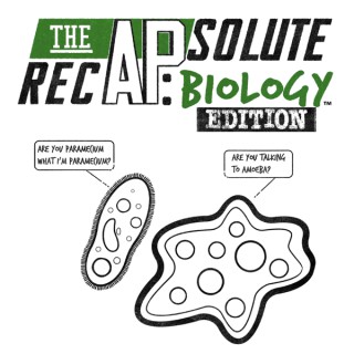
It’s the battle of the nucleic acids in Episode 32! DNA may have all the information, but RNA does all the work. DNA and RNA are made of nucleotide monomers (1:05). DNA and RNA nucleotides differ in their pentose sugar (1:55) and pyrimidine base ( 3:00). DNA forms an antiparallel double helix (3:50) compared to the single strand RNA (5:05), forming mRNA, rRNA and tRNA.The Question of the Day asks (6:44) “Nucleic acids are one of two biological molecules that contain the element nitrogen. What is the other?”Thank you for listening to The APsolute RecAP: Biology Edition!(AP is a registered trademark of the College Board and is not affiliated with The APsolute RecAP. Copyright 2020 - The APsolute RecAP, LLC. All rights reserved.)Website:www.theapsoluterecap.comEMAIL:TheAPsoluteRecAP@gmail.comFollow Us:INSTAGRAMTWITTER
Episode 20 dives into all things transcription and translation with the Central Dogma! Don’t forget that you are what your proteins produce. The mRNA transcript is named after the Latin word for writing (1:50). RNA Polymerase will synthesize mRNA in the 5’-3’ direction, just like DNA polymerase. Do you know Chargaff’s rules? (3:22). The mRNA transcript will need to go through modification as it exits the nuclear pore (3:40). Translation involves mRNA, rRNA and tRNA to form the polypeptide (4:50). Almost all organisms use the same genetic code which supports common ancestry (6:00).The Question of the Day asks (6:48) “Which enzyme copies the viral RNA genome into DNA for the assembly of new viral progeny?”Thank you for listening to The APsolute RecAP: Biology Edition!(AP is a registered trademark of the College Board and is not affiliated with The APsolute RecAP. Copyright 2020 - The APsolute RecAP, LLC. All rights reserved.)Website:www.theapsoluterecap.comEMAIL:TheAPsoluteRecAP@gmail.comFollow Us:INSTAGRAMTWITTER

In this episode of the Epigenetics Podcast, we caught up with Professor Tom Moss from Université Laval in Québec City, Canada to talk about his work on the chromatin structure and dynamics at ribosomal RNA genes. Dr. Tom Moss has been a member of the Department of Molecular Biology, Medical Biochemistry, and Pathology at the Laval University School of Medicine since he was recruited from the University of Portsmouth in the United Kingdom in 1986. Since then he focused on the ribosomal transcription factor Upstream Binding Factor (UBF) and how it regulates the chromatin structure at ribosomal RNA genes (rDNA). UBF binds to the rDNA as a dimer where it leads to six in-phase bends and induces the formation of the ribosomal enhanceosome. This enhanceosome is required for the initial step in formation of an RNA polymerase I initiation complex, and therefore plays an important role in regulating the expression of ribosomal RNA genes. In this Interview, we discuss the function of UBF on the rDNA, how UBF impacts the chromatin landscape at rRNA genes, the role of DNA methylation in this process, and how UBF influences the structure of the nucleolus. References D. Bachvarov, T. Moss (1991) The RNA polymerase I transcription factor xUBF contains 5 tandemly repeated HMG homology boxes (Nucleic Acids Research) DOI: 10.1093/nar/19.9.2331 V. Y. Stefanovsky, D. P. Bazett-Jones, … T. Moss (1996) The DNA supercoiling architecture induced by the transcription factor xUBF requires three of its five HMG-boxes (Nucleic Acids Research) DOI: 10.1093/nar/24.16.3208 V. Y. Stefanovsky, G. Pelletier, … T. Moss (2001) DNA looping in the RNA polymerase I enhancesome is the result of non-cooperative in-phase bending by two UBF molecules (Nucleic Acids Research) DOI: 10.1093/nar/29.15.3241 Elaine Sanij, Jeannine Diesch, … Ross D. Hannan (2015) A novel role for the Pol I transcription factor UBTF in maintaining genome stability through the regulation of highly transcribed Pol II genes (Genome Research) DOI: 10.1101/gr.176115.114 Tom Moss, Jean-Clement Mars, … Marianne Sabourin-Felix (2019) The chromatin landscape of the ribosomal RNA genes in mouse and human (Chromosome Research) DOI: 10.1007/s10577-018-09603-9 Contact Active Motif on Twitter Epigenetics Podcast on Twitter Active Motif on Linked-In Active Motif on Facebook eMail: podcast@activemotif.com

We have a groundbreaking discussion with Prof Philip Hugenholtz co-founder of Microba, a company specialising in Metagenomic Stool Testing using DNA based sequencing to analyse the microbiome. We cover the history and application of DNA based sequencing to classify and identify micro-organisms, using the technology to improve health outcomes on a personalised level, the future prospects for precision medicine, IBD, what a healthy microbiome looks like and much more. Bio: Professor Hugenholtz is a microbiologist who has made contributions in the field of culture-independent analysis of microorganisms. He discovered and characterised numerous previously unrecognised major bacterial and archaeal lineages each with greater evolutionary divergence than animals and plants combined. He has participated in the development and application of metagenomics, the genome-based characterisation of microbiomes, which has revolutionised our understanding of microbial ecology and evolution. He has made several discoveries in environmental and clinical microbiology sometimes overturning decades of misdirected culture-based studies. Topics discussed: Phil’s Origin story Microbial Classification and Morphology The Development of DNA based sequencing technology Carl Woese- 16s rRNA - ribosomal RNA sequencing https://en.wikipedia.org/wiki/Carl_Woese Did we evolve from ancient Archaea? Norm Pace- Application of 16s rRNA sequencing in Ecology https://en.wikipedia.org/wiki/Norman_R._Pace Culture indépendant analytical techniques Craig Venter- Metagenomic Sequencing https://en.wikipedia.org/wiki/Craig_Venter Blueprints for identifying bacteria Development of Sequencing Technology Discovery of Microbes Growing Microbes on plates What is the Greengenes reference database 16s rRNA genes identification vs Whole Genome shotgun sequencing (Metagenomics) Metagenomics, Big Data and health patterns The limitation of 16s rRNA technology- conserved genes Whole Genome Sequencing Resolution ITS gene and Fungal Classification Virus and Parasite Classification The Metagenomic workflow GTDB Database for Whole-genome sequencing Predicting IBD, IBS, Crohns Predicting response to drug response- Depression, Cancer The Gut Microbiome as an early warning system The future of Microbiome What does a healthy cohort’s gut microbiome look like? Discovery of new species Faecalibacterium prausnitzii Anti Inflammatory genes and properties The uniqueness of the gut microbiome Characterising IBD The impact of the immune system Opportunistic Pathogens- Clostridium difficile, Bilophila wadsworthia, Desulfovibrio, Helicobacter pylori Gut Metabolite production by microbes- e.g GABA (a neurotransmitter linked to depression) via KEGG The future of Gut Microbiome Testing Metabolomics Artificial Intelligence and Machine Learning Algorithms for health predictions Diet as a key driver of the microbiome Personalised Diet based on a microbiome profile Prebiotics and Probiotics and Precise Medicine Fibre and Butyrate Balance Phil’s top gut health tip Microbiome changes and improved mental health via diet and exercise Brought to you by: Nourishme Organics- The Gut Health Superstore Check out the Microba Metagenomic Stool Testing and Nutritional Consulting Package for Personalised advice on how to optimise your gut health based on your unique microbiome https://www.nourishmeorganics.com.au/products/gut-explorer-pro-metagenomic-stool-testing-personalised-nutrition-consultation Use code guthealthgurus for 10% off Connect with Prof Phil Hugenholtz Website- https://www.microba.com/ Connect with Kriben Govender: Facebook- https://www.facebook.com/kribengee/ Instagram- https://www.instagram.com/kribengovender/ Youtube- https://www.youtube.com/c/Nourishmeorganics?sub_confirmation=1 Gut Health Gurus Facebook Group: https://www.facebook.com/groups/nourishmeorganics/ Deuterium Depletion Support Facebook Group: https://www.facebook.com/groups/347845406055631/ Download links If you enjoyed this episode and would like to show your support: 1) Please subscribe on Apple Podcasts, give us 5 stars and leave a positive review Instructions: - Click this link https://itunes.apple.com/au/podcast/gut-health-gurus-podcast/id1433882512?mt=2 - Click "View in Itunes" button on the left-hand side - This will open the Itunes app - Click the "Subscribe" button - Click on "Ratings and Reviews" tab - Click on "Write a Review" button Non-Itunes users can leave a Google Review here: https://goo.gl/9aNP0V 2) Subscribe, like and leave a positive comment on Youtube https://www.youtube.com/c/Nourishmeorganics?sub_confirmation=1 3) Share your favourite episode on Facebook, Instagram, and Stories 4) Let your friends and family know about this Podcast by email, text, messenger etc Thank you so much for your support. It means the world to us.

Flávia revisa as principais características dos três tipos de RNA (mRNA, rRNA, tRNA) como preparação e revisão para a sua prova do ENEM. Tenha acesso ao nosso conteúdo online gratuito no link!

What is the best diet? This question remains the million (probably billion) dollar question in health, nutrition and medicine. The winner will likely be the healthy diet is one that prevents the most disease and weight gain. Now there seems to be an approach that has caught the attention of patients and providers internationally and that looking at individualized post-meal glucose response as a guide to one’s individualized diet. And guess what? The researchers say that the post meal glucose response is largely dependent as to what kinds of bugs are growing in our gut. Listen in on today’s episode as we explore the individualized diet and post meal glucose response. Some back ground information. Post meal glucose response is defined classically as the rises in blood glucose measured in ng/dl. Why does it matter? As a biomarker it predicts one’s ability to regulate glucose. Glucose regulated means less inflammation in the body, less risk for heart disease, less risk for diabetes, less risk for kidney disease, less risk for eye disease, and less risk for obesity…you get the picture. Typically in a non-diabetic a fasting blood glucose will range from 70 ng/dl to 99 ng/dl and after eating a carbohydrate containing meal the blood glucose will rise between 100 ng/dl-140ng/dl. This can be influenced by protein and fat content in the food source however it is largely driven by carbohydrate intake. Until researchers started looking to the gut microbiome, the large dietary agencies that be would look at glucose content of foods and make predictions as to how these foods would spike blood sugar. This list is called the glycemic index of foods. Then later they looked at how combinations of foods might spike one’s glucose and this is called glycemic load. Now a group out of the Weizmann Institute developed a test and algorithm of post meal glucose response based on DNA sequencing of the bugs that are growing in human guts. By monitoring glucose response in human subjects, they were able to predict that one’s own signature gut microbiome, can largely influence ones glucose response to food. about our guest Richard Sprague is a heath technology executive, biohacker, citizen scientist and engineer. He is quite the interesting man. He is the CEO of Airdoc (a medical technology company). He has been quite involved with Stool Microbiome testing using 16s rRnA metabolome sequencing. At one point he worked with Ubiome one of the pioneers in stool microbiome testing. He must own the record for the number of times he has tested his own stool microbiome (over 300). With this background he brings an interesting perspective on what is actionable within gut microbiome data. --- Support this podcast: https://anchor.fm/adam-rinde/support

Fakultät für Geowissenschaften - Digitale Hochschulschriften der LMU
Sponges are simple animals that mostly inhabit the marine ecosystem. The role of sponges in the marine ecosystem and the potential of their bioactive compounds for the pharmaceutical industry have already been reviewed. Because of the extensive investigations of sponges within those two disciplines, marine ecology and chemistry, sponges are among the best-studied Metazoa. Likewise, sponges have been selected as animal models for investigating the origin of the multicellularity because sponges have a simple body structure and physiology (e.g., lack of nervous and circulatory organs). Due to their diversity and abundance in the tropics, particularly in the Indo-Pacific, sponges have also attracted taxonomists, systematists and ecologists to assess their diverseness and their phylogenetic and phylogeographic relationships. Resolving those research questions is difficult, because sponges are categorised as comparatively character poor taxa. By using only conservative taxonomy or systematics, the sponge diversity might therefore be underestimated. Inevitably, sponge biologists have to employ molecular methods as additional tools. In this research, molecular tools were used in order to analyse the taxonomy, phylogeny and phylogeographic relationships of selected sponge species. Xestospongia testudinaria & Neopetrosia exigua (Family Petrosiidae, Order Haplosclerida) were selected because of their conspicuousness in the Indo-Pacific coral reef ecosystems, whereby Xestospongia testudinaria is prominently known as the Indo-Pacific giant barrel sponge. Additionally, the order Haplosclerida has been described as an example of sponge order that has been examined systematically for a number of years and displays major discrepancy between morphology and molecular phylogenies. Molecular data suggests that the order needs revision at all taxonomic levels, which is the cause for further conflicts between taxonomists and systematists. In my research I focused mostly on sponge samples that originated from South East Asia or the Indo-Australian Archipelago (IAA). This region represents one of the best-explored marine regions in the Indo-Pacific. The aim of my research is to discover to what extent molecular tools are suitable to detect a phylogenetic signal, a phylogeographical break or a genotypic difference in the two selected sponge taxa. Several markers from the mitochondrial (mtDNA), ribosomal (rRNA) and nuclear (nucDNA) have been utilised. The 3' partition of the cytochrome oxidase subunit 1 (I3-M11 of cox1) from the mtDNA could be used to detect a genetic structure in Xestospongia testudinaria in a geographical narrow scale study of < 200 km2 in Lembeh, North Sulawesi, Indonesia (Chapter 6) and throughout the Indo-Pacific despite limitations in the sample datasets (Chapter 2). In addition, the presence of a species complex in X. testudinaria was detected with the aid of phylogenetic reconstructions from a concatenation of mtDNA sequences (I3-M11 of cox1 and the Adenosine Triphosphate Synthase F0 subunit 6 / ATP6), and a nucDNA marker, the Adenosine Triphosphate Synthase β subunit intron (ATPS-β intron) (Chapter 6). At the same time, the presence of a species complex in X. testudinaria was recognised in a broader scale study of the Indo-Australian Archipleago (IAA) (Chapter 3). As a result, selected mtDNA and nucDNA markers in this thesis are useful for the investigation of the taxonomical status and phylogeographical relationships of X. testudinaria. A phylogeographical break in the IAA region due to the Pleistocene low sea level and Holocene recolonisation events (Chapter 3) could not be recovered among X. testudinaria in a phylogeographical analysis. Similarly, overlapping I3-M11 cox1 haplotypes between X. testudinaria, X. muta and X. bergquistia were recovered. This might be due to the presence of ancient polymorphisms on the barrel sponge mtDNA markers. Molecular tools are also used to help identifying my second selected sponge species (Chapter 4). The use of selected cox2 mtDNA and 28S rRNA markers contributed significantly to the identification of. Neopetrosia exigua used to be a congeneric of X. testudinaria. During my examinations of self-collected and holotype specimens I discovered that the species named N. exigua bears a wrong name. For this reason, a taxonomical revision is suggested and, more importantly, according to my findings and the principle of priority in the ICZN (International Code of Zoological Nomenclature) I use the species name ‘chaliniformis’ instead of the species name ‘exigua’. Furthermore, the use of selected nucDNA marker, the Lysidyl Aminoacyl Transfer RNA Synthetase (LTRS) intron, also contributes to the detection of phylogeographical breaks in N. chaliniformis of the IAA (Chapter 5). In a nutshell, the success of unravelling sponge taxonomies, phylogenies, and phylogeographic relationships always depends on the suitability of the utilised molecular markers and the significance of environmental influences on the sponges. Haplosclerid sponges possess limited morphological features. These hurdles create several problems, e.g. difficulties with taxa delimitation and unresolved phylogeography relationships. Even though the application of molecular techniques generated some limitations and obstacles in these studies, it has already contributed significantly to a better understanding of the phylogenies, phylogeographic relationships and taxonomical problems of X. testudinaria and N.chaliniformis, the species I selected for my research.

Fakultät für Chemie und Pharmazie - Digitale Hochschulschriften der LMU - Teil 05/06
To study the mechanisms of membrane protein insertion we established a protocol that allows isolation of in vivo assembled ribosome nascent chain complexes (RNCs) from E. coli in high yield and quality. To investigate the interaction of SecY with a translating ribosome, model membrane proteins of different length and topology were over-expressed and the respective RNCs were isolated under mild conditions to allow co-purification of the SecY complex. Analysis of the interaction of RNCs with SecY in vivo suggested that, as expected, a tight engagement of the ribosome and SecY is only established for nascent chains that are translocated co-translationally. We observed that SecY and the RNC do not form a stable complex at the moment of hydrophobic transmembrane segments inserting in the translocon. However, a stable engagement of the RNC with SecY was observed, when inserting a transmembrane segment with a type II topology into SecY followed by a hydrophilic loop of a certain length which allows the isolation of this complex. That suggested a dual binding mode of tight and loose coupling of SecY to the translating ribosome dependent on the nature of the nascent substrate. We present the first three dimensional structure of an in vivo assembled, tightly coupled polytopic RNC-SecYE complex at 7.2 Å solved by cryo-EM and single particle reconstruction. A molecular model based on the cryo-EM structure reveals that SecYE could be trapped in a post-insertion state, with the two substrate helices interacting with the periphery of SecY, while still translocating the hydrophilic loop. The lateral gate of SecY remains in a ‘pre-opened’ conformation during the translocation of the hydrophilic loop. The interaction sites of SecY with the ribosome were found as described. Remarkably, we could also reveal an interaction of helix 59 in the ribosome with nascent membrane protein via positively charged residues in the first cytoplasmic loop of the substrate. It is tempting to speculate that this interaction contributes to the positive inside rule. Though, we provided an unprecedented snapshot of an inserting polytopic membrane protein, the exact path of the nascent chain and the molecular mechanism of the actual insertion could not be solved so far. Expression of the E. coli tryptophanase (TnaA) operon is triggered by ribosome stalling during translation of the upstream TnaC leader peptide. Notably, this stalling is strictly dependent on the presence of tryptophan that acts in a hitherto unknown way. Here, we present a cryo-EM reconstruction of the stalled nascent TnaC leader peptide in the ribosomal exit tunnel. The structure of the TnaC-stalled ribosome was solved to an average resolution of 3.8 Å by cryo-EM and single particle analysis. It reveals the conformation of the silenced peptidyl-transferase center as well as the exact path of the stalled nascent peptide and its contacts in detail. Furthermore, we clearly resolve not a single but two free tryptophan molecules in the ribosomal exit tunnel. The nascent TnaC peptide chain together with distinct rRNA bases in the ribosomal exit tunnel creates two hydrophobic binding pockets for the tryptophan coordination. One tryptophan molecule is coordinated by V20 and I19 of TnaC and interacts with U2586 of the rRNA, the second tryptophan is bound between I19 and I15 in the area of A2058 and A2059 of the rRNA. Interestingly, the latter is also the binding platform for macrolide antibiotics. Engagement of L-Trp in these composite binding pockets leads to subtle conformational changes in residues of the ribosomal tunnel wall that are translated to the PTC eventually resulting in silencing by stabilizing the conformations of the conserved nucleotides A2602 and U2585. These conformations of the two nucleotides in the PTC are incompatible with the correct accommodation of the GGQ motive of release factor 2, thus inhibiting the peptide release.

James M. Guyette is President and Chief Executive Officer of Rolls-Royce North America Inc. (RRNA). (December 18, 2013)
Fakultät für Biologie - Digitale Hochschulschriften der LMU - Teil 04/06
Tue, 12 Jun 2012 12:00:00 +0100 https://edoc.ub.uni-muenchen.de/14511/ https://edoc.ub.uni-muenchen.de/14511/1/Feistel_Susanne.pdf Feistel, Susanne ddc:570, ddc:500, Fakultät für Biologie

Medizinische Fakultät - Digitale Hochschulschriften der LMU - Teil 14/19
Thu, 8 Mar 2012 12:00:00 +0100 https://edoc.ub.uni-muenchen.de/14136/ https://edoc.ub.uni-muenchen.de/14136/1/Freitag_Martin.pdf Freitag, Martin
Fakultät für Chemie und Pharmazie - Digitale Hochschulschriften der LMU - Teil 04/06
Wed, 25 Jan 2012 12:00:00 +0100 https://edoc.ub.uni-muenchen.de/14677/ https://edoc.ub.uni-muenchen.de/14677/1/Jennebach_Stefan.pdf Jennebach, Stefan ddc:540, ddc:500, Fakultät für Chemie und Pharmazie
Fakultät für Chemie und Pharmazie - Digitale Hochschulschriften der LMU - Teil 04/06
Eukaryotic nuclear transcription is carried out by three different Polymerases (Pol), Pol I, Pol II and Pol III. Among these, Pol I is dedicated to transcription of the rRNA, which is the first step of ribosome biogenesis, and cell growth is regulated during Pol I transcription initiation by the conserved factor Rrn3/TIF-IA in yeast/human. A wealth of structural information is available on Pol II and its general transcription factors (GTFs). Recently, also the architectures of Pol I and Pol III have been described by electron microscopy and the additional subunits that are specific to Pol I and Pol III have been identified as orthologs of the Pol II transcription factors TFIIF and TFIIE. Nevertheless, we still lack information about the architecture of the Pol I initiation complex and structural data is missing explaining the regulation of Pol I initiation mediated by its central transcription initiation factor Rrn3. The Rrn3 structure solved in this study reveals a unique HEAT repeat fold and indicates dimerization of Rrn3 in solution. However, the Rrn3-dimer is disrupted upon Pol I binding. The Rrn3 structure further displays a surface serine patch. Phosphorylation of this patch represses human Pol I transcription (Mayer et al, 2005; Mayer et al, 2004), and a phospho-mimetic patch mutation prevents Rrn3 binding to Pol I in vitro, and reduces S. cerevisiae cell growth and Pol I gene occupancy in vivo. This demonstrates a conserved regulation mechanism of the Pol I-Rrn3 interaction. Crosslinking indicates that Rrn3 does not only interact with Pol I subunits A43/14, but the interface further extends past the RNA exit tunnel and dock domain to AC40/19. The corresponding region of Pol II binds the Mediator head (Soutourina et al., 2011) that co-operates with TFIIB (Baek et al, 2006). Consistent with this, the Rrn3 binding partner, core factor subunit Rrn7, is predicted to be a TFIIB homologue. Taken together, our results provide the molecular basis of Rrn3-regulated Pol I initiation and cell growth and indicate a universally conserved architecture of eukaryotic transcription initiation complexes.
Fakultät für Chemie und Pharmazie - Digitale Hochschulschriften der LMU - Teil 04/06
Topic I Molecular basis of RNA polymerase III transcription repression by Maf1 RNA polymerase III (RNAP III) is a conserved 17-subunit enzyme that transcribes genes encoding short untranslated RNAs such as transfer RNAs (tRNAs) and 5S ribosomal RNA (rRNA). These genes are essential and involved in fundamental processes like protein biogenesis; hence RNAP III activity needs to be tightly regulated. RNAP III is repressed upon stress and this is regulated by Maf1, a protein conserved from yeast to humans. Many stress pathways were shown to converge on Maf1 and result in its phosphorylation, followed by its nuclear import and eventual repression of RNAP III activity. However, the molecular mechanisms of this repression activity were not known at the beginning of these studies. This work establishes the mechanism of RNAP III specific transcription repression by Maf1. The crystal structure of Maf1 was solved. It has a globular fold with surface accessible NLS sequences, which sheds new light on already published results and explains how stress-induced phopshorylation leads to import of Maf1 into the nucleus. Additionally, cryo EM studies and competition assays show that Maf1 binds RNAP III at its clamp domain and thereby induces structural rearrangements of RNAP III, which inhibits the interaction with Brf1, a subunit of the transcription initiation factor TFIIIB. This specifically impairs recruitment of RNAP III to its promoters and implies that Maf1 is a repressor of transcription initiation. Competition and transcription assays show that Maf1 also binds RNAP III that is engaged in transcription, leaving RNAP III activity intact but preventing re-initiation. Topic II Structure of human mitochondrial RNA polymerase The nuclear-encoded human mitochondrial RNAP (mitoRNAP) transcribes the mitochondrial genome, which encodes rRNA, tRNAs and mRNAs. MitoRNAP is a single subunit (ss) polymerase, related to T7 bacteriophage and chloroplast polymerases. All share a conserved C-terminal core, whereas the N-terminal parts of mitoRNAP do not show any homology to other ss RNAPs. Unlike phage RNAPs, which are self-sufficient, human mitoRNAP needs two essential transcription factors for initiation, TFAM and TFB2M. Both of these factors are likely to control the major steps of transcription initiation, promoter binding and melting. Thus human mitoRNAP has evolved a different mechanism for transcription initiation and exhibits a unique transcription system. Structural studies thus far concentrated on the nuclear enzymes or phage RNAPs, whereas the structure of mitochondrial RNA polymerase remained unknown. The structural organization of human mitoRNAP and the molecular mechanisms of promoter recognition, binding and melting were subject of interest in these studies. In this work the crystal structure of human mitoRNAP was solved at 2.4 Å resolution and reveals a T7-like C-terminal catalytic domain, a N-terminal domain that remotely resembles the T7 promoter-binding domain (PBD), a novel pentatricopeptide repeat (PPR) domain, and a flexible N-terminal extension. MitoRNAP specific adaptions in the N-terminus include the sequestering of one of the key promoter binding elements in T7 RNAP, the AT-rich recognition loop, by the PPR domain. This sequestration and repositioning of the N-terminal domain explain the need for the additional initiation factor TFAM. The highly conserved active site within the C-terminal core was observed to bind a sulphate ion, a well known phosphate mimic, and thereby suggests conserved substrate binding and selection mechanisms between ss RNAPs. However, conformational changes of the active site were observed due to a movement of the adjacent fingers subdomain. The structure reveals a clenching of the active site by a repositioned fingers subdomain and an alternative position of the intercalating -hairpin. This explains why the conserved transcription factor TFB2M is required for promoter melting and initiation. A model of the mitochondrial initiation complex was build to further explore the initiation mechanism, and to rationalize the available biochemical and genetic data. The structure of mitoRNAP shows how this enzyme uses mechanisms for transcription initiation that differ from those used by phage and cellular RNAPs, and which may have enabled regulation of mitochondrial gene transcription and adaptation of mitochondrial function to changes in the environment.
Fakultät für Biologie - Digitale Hochschulschriften der LMU - Teil 04/06
Mon, 18 Jul 2011 12:00:00 +0100 https://edoc.ub.uni-muenchen.de/16092/ https://edoc.ub.uni-muenchen.de/16092/1/Hochstatter_Julia.pdf Hochstatter, Julia ddc:570, ddc:500, Fakultät für
Fakultät für Biologie - Digitale Hochschulschriften der LMU - Teil 04/06
Die Biogenese eukaryotischer Ribosomen ist ein streng kontrollierter, dynamischer Prozess, der ein komplexes räumliches und zeitliches Zusammenspiel vieler verschiedener Proteine erfordert. Dabei wird zunächst ribosomale DNA mit Hilfe der RNA-Polymerase I im Nukleolus transkribiert. Das daraus resultierende rRNA-Vorläufer-Molekül wird anschließend umfassend prozessiert und modifiziert. Gleichzeitig assemblieren ribosomale Proteine mit der reifenden rRNA, um präribosomale Partikel zu bilden, die für weitere Reifungsschritte ins Nukleoplasma und Cytoplasma transportiert werden. Zum Verständnis des ribosomalen Reifungsprozesses haben bislang vor allem genetische und biochemische Studien in der Bäckerhefe beigetragen. In Säugerzellen sind dagegen die Komponenten der Ribosomenbiogenese wenig charakterisiert, und insbesondere unser Wissen über die Regulationsmechanismen ist lückenhaft. Die posttranslationale Modifikation mit dem ubiquitinähnlichen SUMO-Protein reguliert eine Vielzahl wichtiger zellulärer Prozesse. SUMO-spezifische Isopeptidasen der SENP-Familie katalysieren die Abspaltung von SUMO von Zielproteinen und kontrollieren damit das Gleichgewicht zwischen Modifikation und Demodifikation. Vorarbeiten zu dieser Arbeit haben eine entscheidende Rolle für die SUMO-Isopeptidase SENP3 während der nukleolären Schritte der Ribosomenbiogenese gezeigt. Allerdings waren die Substrate von SENP3 bei diesem Prozess weitgehend unbekannt. Im Rahmen dieser Arbeit wurde ein SENP3-assoziierter Proteinkomplex bestehend aus den Komponenten PELP1, TEX10 und WDR18 aufgereinigt. Als weitere Bindungspartner von PELP1 wurden außerdem MDN1 und LAS1L identifiziert. Durch RNAi-vermittelte Depletionsexperimente in Zellkultur konnte gezeigt werden, dass PELP1, TEX10 und WDR18, sowie die assoziierten Proteine MDN1 und LAS1L, ebenso wie SENP3 für die Reifung der 28S rRNA und den nukleolären Export der großen ribosomalen Untereinheit erforderlich sind. PELP1 und LAS1L wurden als SENP3-sensitive SUMOSubstrate charakterisiert. Darüberhinaus konnte gezeigt werden, dass das Gleichgewicht zwischen Sumoylierung und Desumoylierung die subnukleäre Lokalisierung des Komplexes kontrolliert. Sumoylierung führt zum Ausschluss aus dem Nukleolus, während Desumoylierung die nukleoläre Kompartimentierung fördert. Zusammengefasst zeigen die Ergebnisse der vorliegenden Arbeit, dass der Komplex aus den Proteinen PELP1, TEX10 und WDR18 die Ribosomenbiogenese reguliert. Außerdem deuten sie darauf hin, dass dessen SUMO-abhängige subzelluläre Verteilung Ablauf und Koordination der Ribosomenreifung kontrolliert.
Fakultät für Chemie und Pharmazie - Digitale Hochschulschriften der LMU - Teil 04/06
Ribosomes are macromolecular protein-RNA complexes translating mRNA into protein. To date, crystal structures are available for the bacterial 30S and archaeal 50S subunits, as well as the complete bacterial 70S ribosomes. Eukaryotic ribosomes are much more complex in terms of ribosomal RNA and proteins. However, to date high-resolution crystal structures of eukaryotic ribosomes or ribosomal subunits are lacking. In order to build reliable models for the eukaryotic rRNA, we developed an approach for large scale homology and de novo modeling of RNA and subsequent exible tting into high-resolution cryo-EM density maps. Using this approach we built a model of the T. aestivum and the S. cerevisiae ribosome based on available cryo-EM maps at 5.5 Å and 6.1 Å resolution, respectively. The model comprises of 98% of the eukaryotic rRNA including all 21 RNA expansion segments (ES) and structurally six variable regions. Further, we were able to localize 74/80 (92.5%) of the ribosomal proteins. The model reveals unique ES-ES and r-protein-ES interactions, providing new insight into the structure and evolution of the eukaryotic ribosome. Moreover, the model was used for analyzing functional ribosomal complexes, i.e. the characterization of dierent nascent polypeptide chains within the ribosomal tunnel, intermediates of protein translocation as well as mRNA quality control.
Fakultät für Chemie und Pharmazie - Digitale Hochschulschriften der LMU - Teil 04/06
Protein biosynthesis, the translation of the genetic code into polypeptides, occurs on ribonucleoprotein particles called ribosomes. Although X-ray structures of bacterial ribosomes are available, high-resolution structures of eukaryotic 80S ribosomes are lacking. Using cryo-electron microscopy and single-particle reconstruction we have determined the structure of a translating plant (Triticum aestivum) 80S ribosome at 5.5 Å resolution. This map, together with a 6.1 Å map of a Saccharomyces cerevisiae 80S ribosome, has enabled us to model ~98 % of the rRNA and localize 74/80 (92.5 %) of the ribosomal proteins, encompassing 11 archaeal/eukaryote-specific small subunit proteins as well as the complete complement of the ribosomal proteins of the eukaryotic large subunit. Near-complete atomic models of the 80S ribosome provide insights into the structure, function and evolution of the eukaryotic translational apparatus.
Fakultät für Biologie - Digitale Hochschulschriften der LMU - Teil 03/06
Plant beneficial microorganisms, such as arbuscular mycorrhiza fungi (AMF), increasingly attract scientific and agronomic attention due to their capacity to increase nutrient accessibility for plants and to reduce inorganic fertilizer requirements. AMF are thought to form symbioses with most land plants, obtaining carbon from the autotrophic host whilst enhancing uptake of poorly available nutrients. The species of AMF are mainly identified by spore morphology, which is time consuming, requires expertise and is rarely applicable to AMF identification in roots. Molecular tools such as analysis of standardized DNA fragment sequences may allow the recognition of species through a ‘DNA barcode’, which may partly overcome this problem. The focus of this study was to evaluate different regions of widely used rDNA repeats for their use as DNA barcodes for AMF including the small subunit rRNA gene (SSU), the internal transcribed spacer (ITS) and the large subunit rRNA gene (LSU). Closely related species in the genus Ambispora, members of which have dimorphic spores, could not be separated by analysis of the SSU region, but of the ITS region. Consequently, the SSU was not used for subsequent analysis, but a DNA fragment covering a small part of the SSU, the entire ITS region and about 800 bp of the LSU (SSUmCf-LSUmBr fragment) was analysed, providing phylogenetic resolution to species. New AMF specific primers for these potential barcoding regions were developed and can be applied, without amplification of non-target organisms, for AMF species determination, including identification from field and root samples. Analyses based on the application of the SSUmCf-LSUmBr fragment showed that the widely used AMF model organism Glomus sp. DAOM197198 (formerly called Glomus intraradices) is not conspecific with Gl. intraradices. The SSUmCf-LSUmBr fragment clearly provides a much higher species resolution capacity when compared with the formerly preferred ITS and LSU regions. Further study of several groups of AMF species using different regions of the SSUmCf-LSUmBr fragment revealed that only the complete SSUmCf-LSUmBr fragment allowed separation of all analysed species. Based on these results, an extended DNA barcode covering the ITS region and parts of the LSU region is suggested as a DNA barcode for AMF. The complete SSUmCf-LSUmBr fragment sequences can serve as a database backbone for also using smaller rDNA fragments as barcodes. Although the smallest fragment (approximately 400 bp) analysed in this study was not able to discriminate among AMF species completely, such short regions covering the ITS2 or LSU D2 regions, respectively, would most likely be suitable for community analyses with 454 GS-FLX Titanium sequencing, providing that the analyses is based on the longer DNA sequences.
Fakultät für Biologie - Digitale Hochschulschriften der LMU - Teil 03/06
Während der Zellproliferation müssen Zellwachstum und Zellteilung koordiniert werden. Die Kopplung erfolgt in der Hefe durch einen Komplex aus Nop7p, Erb1p und Ytm1p, der sowohl an der Ribosomenbiogenese als auch an der Kontrolle der DNA-Replikation beteiligt ist. Die homologen Proteine Pes1, Bop1 und WDR12 werden in Säugern von Zielgenen des Transkriptionsfaktors c-Myc, einem zellulären Onkoprotein, kodiert. In dieser Arbeit wurde die Existenz eines evolutionär konservierten Komplexes aus Pes1, Bop1 und WDR12 (PeBoW-Komplex) in Säugern belegt. Dabei wurde gezeigt, dass Bop1 als zentrales Protein des Komplexes agiert und die Interaktion von Pes1 und WDR12 vermittelt. Die Integrität des Komplexes ist wesentlich für seine Funktion. Die Depletion einzelner Komponenten sowie die Überexpression des integrierenden Proteins Bop1 hemmen die Reifung der Vorläufer-rRNA der großen ribosomalen Untereinheit sowie die Proliferation der Zellen. Bop1-Überexpression führt zur Ausbildung von zwei Subkomplexen aus Bop1 und Pes1 bzw. Bop1 und WDR12. Während der Bop1/Pes1-Subkomplex als Teil der pre-Ribosomen im Nukleolus lokalisiert, wird WDR12 durch Bop1-Überexpression im Zytoplasma gehalten und fehlt im Nukleolus zur Ausbildung eines funktionellen PeBoW-Komplexes. Pes1 und WDR12 können unabhängig in den Nukleolus translozieren, während Bop1 dafür die Interaktion mit Pes1 benötigt. Untersuchungen zur Stabilität der einzelnen PeBoW-Komponenten zeigten, dass monomeres Bop1 extrem instabil ist, durch Inkorporation in den PeBoW-Komplex aber vor Abbau geschützt wird. Möglicherweise werden hierdurch interne PEST-Sequenzen in Bop1 maskiert. Die Menge an Bop1 ist somit abhängig von der Anwesenheit von Pes1 und WDR12. Die gegenseitige Abhängigkeit der Stabilität aller drei PeBoW-Komponenten konnte in weitergehenden Experimenten gezeigt werden. Schließlich wurde untersucht, ob der PeBoW-Komplex die Ribosomenbiogenese mit der DNA-Replikation über Interaktion mit dem ORC-Komplex, wie in der Hefe beschrieben, koordiniert. Mit Hilfe der BiFC-Methode konnte eine Interaktion von Pes1 mit Orc6, eines Faktors des ORC-Komplexes, gezeigt werden. Die koordinierende Funktion des PeBoW-Komplexes für Zellwachstum und Zellproliferation scheint von der Hefe bis zum Menschen stark konserviert zu sein.
Fakultät für Chemie und Pharmazie - Digitale Hochschulschriften der LMU - Teil 02/06
Synthesis of ribosomal RNA by RNA polymerase (Pol) I is the first step in ribosome biogenesis and a regulatory switch in eukaryotic cell growth. In this thesis a reproducible large-scale purification protocol for Pol I from S. cerevisiae could be developed. Crystals were obtained, diffraction to < 4 Å could be recorded, however, the enormously complex non-crystallographic symmetry impeded structure solution. Switching to cryo-electron microscopy, the structure of the complete 14-subunit enzyme could be solved to 12 Å resolution, a homology model for the core enzyme could be generated, and the crystal structure of the subcomplex A14/43 could be solved. In the resulting hybrid structure of Pol I, A14/43, the clamp, and the dock domain contribute to a unique surface interacting with promoter-specific initiation factors. The Pol I-specific subunits A49 and A34.5 form a heterodimer near the enzyme funnel that acts as a built-in elongation factor, and is related to the Pol II-associated factor TFIIF. In contrast to Pol II, Pol I has a strong intrinsic 3’-RNA cleavage activity, which requires the C-terminal domain of subunit A12.2, and apparently enables rRNA proofreading and 3’-end trimming.
Fakultät für Biologie - Digitale Hochschulschriften der LMU - Teil 03/06
The hallmark of embryonic stem (ES) cells is their ability for self-renewal (capability of unlimited cell division without the loss of pluripotency) as well as for differentiation into all cell types of the adult organism. One factor supposed to be involved in self-renewal is the rapid proliferation rate of ES cells, which is coupled to an unusual cell cycle distribution with the majority of cells in S-phase and a very short G1-phase. This is linked to the lack of a functional G1/S-phase checkpoint, which allows the cells to enter the S-phase almost directly after mitosis. Generally, cells have to closely coordinate growth and cell cycle progression during proliferation to prevent premature division. One important factor for cell growth is ribosome biogenesis. In mature cells, disruptions in ribosome biogenesis are directly linked to the cell cycle machinery by a p53-dependent activation of the G1/S-phase checkpoint, leading to an arrest of cells in G1-phase. During this work, the function of the proteins Pes1, Bop1 and WDR12, which were shown previously to be involved in ribosome biogenesis of mature cell lines, was investigated in mouse ES cells. Moreover, a putative crosstalk between ribosome biogenesis and proliferation of ES cells was assessed. A high expression of Pes1, Bop1 and WDR12 was observed in ES cells, which strongly decreased during in vitro differentiation. Localization of the proteins was predominantly nucleolar and the formation of a stable complex (PeBoW-complex), including all three proteins, was experimentally validated in mature mouse cells as well as in mouse ES cells. The function and stability of the proteins seems to be dependent on incorporation into the PeBOW-complex, as protein levels were interdependent on each other and no free, non-incorporated proteins were observed, except for WDR12. According to their nucleolar localization, depletion of Pes1 and Bop1 were shown to inhibit maturation of the 28S rRNA and thereby the large 60S ribosomal subunit. Further, impaired proliferation of ES cells was observed. Thus, the PeBoW-complex seems to be an essential factor for the rapid proliferation of ES cells and might therefore also be involved in self-renewal. However, first results suggest that the complex is not directly involved in the maintenance of pluripotency. No changes in the expression levels of pluripotency-genes like Nanog, KLF4 and Sox2 were observed. Moreover, alkaline phosphatase activity was equally detectable after depletion of Pes1 or Bop1 and no morphological changes within the ES cell colonies were observed. Impaired ribosome biogenesis is known to activate a p53-dependent checkpoint in mature cell lines, which leads to an arrest of cells in G1-phase. Treatment of mouse NIH3T3 cells with 5FU, a potent inhibitor of rRNA maturation, confirmed an activation of this checkpoint, leading to weak induction of the tumor suppressor p53, induction of the Cdk-inhibitor p21, an increase in active, hypo-phosphorylated Rb, and to accumulation of cells in the G1- and S-phase with an increase of cells in G1-phase. In contrast, ES cells showed strong induction of p53, but no induction of its target gene p21. The overall levels of Rb were strongly induced, but the ratio between inactive, hyper-phosphorylated Rb and active, hypo-phosphorylated Rb was not changed towards the active form. These results were observed upon 5FU treatment and upon depletion of Pes1 or Bop1. Hence, ribosomal stress does not lead to checkpoint activation via the p53-p21-Rb pathway in ES cells. Moreover, no robust accumulation of cells in G1-phase was observed. 5FU treated ES cells showed an accumulation of cells in S-phase instead. Whether this effect is regulated by the induced p53 needs further investigation. Overall, the results suggest that ES cells use different mechanisms as mature cells to coordinate their proliferation rate with ribosome biogenesis.

Medizinische Fakultät - Digitale Hochschulschriften der LMU - Teil 07/19
Thu, 22 Nov 2007 12:00:00 +0100 https://edoc.ub.uni-muenchen.de/7738/ https://edoc.ub.uni-muenchen.de/7738/1/Schmidt_Caroline.pdf Schmidt, Caroline
Fakultät für Chemie und Pharmazie - Digitale Hochschulschriften der LMU - Teil 02/06
Regulation of gene expression takes place in the nucleus in a highly structured and condensed nucleoprotein environment, called chromatin (Felsenfeld and Groudine, 2003; Khorasanizadeh, 2004; Vaquero et al., 2003). A broad group of factors regulates the properties of chromatin; e.g. by covalently modifying histones and / or by ATP-dependent chromatin remodeling, thereby allowing or preventing gene expression. The mammalian genome contains hundreds of gene copies encoding precursor ribosomal RNA and the transcription of these genes is highly regulated with respect to cellular metabolism (Grummt, 2003). However, even in actively growing cells, only a subset of the rRNA genes are actively transcribed, exhibiting an accessible chromatin conformation (Conconi et al., 1989). In a chromatin context, the activation of rDNA genes involves the transcription termination factor TTF-I (Längst et al., 1998; Längst et al., 1997a). However, the silenced rDNA gene fraction remains in an inaccessible heterochromatic state throughout the cell cycle (Conconi et al., 1989). Until recently, the onset of silencing and the mechanisms that maintain the inactive state of rRNA genes were less understood. Recent studies, including the work presented in this thesis, provide insights into the molecular mechanism of ribosomal RNA gene silencing (Lawrence et al., 2004; Németh et al., 2004; Santoro and Grummt, 2001; Santoro et al., 2002; Strohner et al., 2004; Zhou et al., 2002). Accumulating evidence indicates that the combined action of chromatin modifying mechanisms such as chromatin remodeling, histone modification and DNA methylation contribute to the process of rRNA gene silencing. Here I present data demonstrating an active role of the chromatin remodeling complex NoRC in rDNA gene silencing and propose dual functions of TTF-I in rDNA regulation in chromatin, namely involvement in both activation and silencing of rDNA transcription. 4.1 NoRC, a novel chromatin remodeling complex In this doctoral study, a novel protein complex, composed of the nucleolar protein Tip5 and the ATPase Snf2h, was purified using convential chromatography and affinity purification methods. A detailed chromatin remodeling analysis revealed that this complex is able to induce mononucleosome movement in an ATP and histone H4 tail dependent fashion. Finally, this Tip5-Snf2h complex was termed NoRC (nucleolar remodeling complex), a novel member of the ISWI family of ATP-dependent chromatin remodeling complexes (Strohner et al., 2001). To dissect its functions, the NoRC complex was reconstituted from its recombinant subunits Tip5 and Snf2h, using the baculo virus driven expression system. Reconstitution confirmed the direct interaction between Tip5 and Snf2h. Furthermore, recombinant and cellular NoRC display similar sizes in gel filtration columns. Recombinant NoRC exhibits chromatin stimulated ATPase activity and mobilizes nucleosomes in an energy-dependent manner. Both activities are histone H4 tail dependent. NoRC and its subunits Tip5 and Snf2h were compared in different DNA / Nucleosome binding assays. NoRC shows preferred binding to structured (bent) DNA, e.g. a region within the mouse rDNA promoter, and interacts with mononucleosomes in electrophoretic mobility shift assays (EMSA). While no stable interaction with core nucleosomes could be detected in EMSA, ATPase assays and DNase I protection assays noticeably pinpointed to NoRC / nucleosome interactions with both nucleosomal and protruding linker DNA. 4.2 NoRC specifically represses rDNA transcription in chromatin The functional consequences of the Tip5 / TTF-I interaction were assessed and the influence on chromatin structure of the rDNA promoter in an in vitro system was determined. Tip5 in NoRC interacts with the N-terminal part of full length TTF-I and unmasks its DNA binding site. This interaction is required both for binding of TTF-I to its promoter-proximal target site and for the recruitment of NoRC to the promoter in chromatin. After association with the rDNA promoter, NoRC alters the position of the promoter-bound nucleosome. To elucidate a potential role of NoRC in rDNA transcriptional regulation, we used an in vitro transcription system with an rDNA minigene reconstituted into chromatin. These studies revealed a specific function for NoRC in rDNA transcriptional repression on chromatin templates. In contrast, NoRC had no effect on DNA transcription. Transcription experiments were then performed with chromatin templates reconstituted from recombinant histones lacking individual histone tails. The results indicate that NoRC-mediated rDNA gene repression is dependent on the histone H4 tail, suggesting an involvement of chromatin remodeling. Further transcription experiments revealed that NoRC-mediated repression occurs prior to preinitiation complex formation and does not affect activated rDNA genes. NoRC stably associates with the silenced gene, and these early steps of rDNA repression do not depend on DNA and histone modifications (Strohner et al., 2004). NoRC showed preferred binding to a structured (bent) region within the mouse rDNA promoter. Methylation of a single CpG dinucleotide within this region abrogated rDNA transcription in chromatin (Santoro and Grummt, 2001), but did not influence DNA binding of NoRC. Furthermore, nucleosomal DNA is less methylated than free DNA, but chromatin remodeling enhances methylation. The results suggest an important role for the chromatin remodeling complex NoRC in the establishment of rDNA silencing. NoRC then contributes to maintenance of the silenced state throughout the cell cycle by interacting with DNA and histone modifying enzymes. Transcriptional repression by chromatin remodeling factors seems to be a common mechanism to stably inhibit gene expression.

Medizinische Fakultät - Digitale Hochschulschriften der LMU - Teil 02/19
RNA (rRNA), die eine wichtige, primär funktionale Rolle in der Zellphysiologie einnimmt. Die Gene dieser rRNA lassen sich in hochkonservierte und hochpolymorphe Bereiche unterscheiden. Während in den konservierten Regionen kaum Mutationen zu finden sind, werden in den polymorphen Abschnitten je nach Grad der Verwandtschaft große Sequenzunterschiede zwischen den verschiedenen Tierarten beobachtet. Individuen einer Art zeigen diese Unterschiede nicht. Somit können diese artspezifischen Abweichungen dazu genutzt werden, biologische Materialien unbekannter Herkunft einer bestimmten Tierart zuzuordnen. Mit der Amplifikation und anschließenden Sequenzierung eines Bereiches innerhalb des mitochondrialen 12S-Gens sowie der Auswertung bereits publizierter Sequenzen gelingt die Identifizierung einer Spezies anhand von 20 – 25 Basen. Dazu kann neben der etablierten Methode der Sequenzierung nach Sanger auch die Technik der Pyrosequenzierung genutzt werden. Die Ergebnisse zeigen die Möglichkeit der Identifizierung verschiedener Arten durch die Analyse kurzer Fragmente ihrer 12S-Gen-Sequenz. Dazu reicht die Amplifikation des gewünschten Fragmentes mit Hilfe eines Primerpaares. Für degradierte DNA wurde ein alternativer Rückprimer getestet, der die Amplifikation eines kürzeren Sequenzabschnittes ermöglicht. Es wurden insgesamt 91 Proben verschiedener Tiere analysiert, die sich aus 8 Säugetier-Arten, 2 Fisch-Arten und 3 Vogel-Arten zusammen setzten. Zudem kann und wird die Methode bereits bei Fragestellungen in der forensischen Routine angewendet.

Fakultät für Biologie - Digitale Hochschulschriften der LMU - Teil 01/06
The aim of this study was to increase the understanding of diversity and activity of dominant bacterial populations in the rhizospheres of three economically important grain legumes (Vicia faba, Lupinus albus and Pisum sativum). A cultivation-independent approach was employed to achieve this aim bearing in mind the limitation of cultivation-dependent technique that only 10% of bacteria present in rhizosphere can be cultured. PCR amplification of 16S rDNA and subsequent separation of the amplicons by DGGE was used in an initial screening of replicates for experimental variation and for the first characterization of bacterial community composition of the three rhizospheres under study. Specific profiles generated by the three legumes, derived by both 16S rDNA and rRNA, emphasized the need to perform detailed analysis of the communities present in these rhizospheres. Clone libraries for PCR and RT-PCR products were generated for representative samples of all the three legumes. Firmicutes were found to be the most dominant in all the legumes, both in DNA- and RNA-derived libraries, indicating them to be the most active group as well. A plant-dependent rhizosphere effect was reflected by the absence of ?-subdivision members in Pisum and ?-subdivision members of proteobacteria in Vicia rhizosphere. High numbers of as yet unclassified bacteria were also obtained. With this experimental set-up, using the same soil material but three different legumes and a uniform inoculation with Rhizobium sp., it became evident that plant roots influence the development of bacterial communities in the rhizosphere in a plant-specific manner. The extent of the rhizosphere effect could vary in natural field conditions as the present study was performed under controlled conditions in green house using soil from agricultural site. Extraction and analysis of rRNA has enabled identification of active taxa in the present study. Fingerprints were obtained for total RNA using two different primers. The profiles generated revealed marked differences between the three rhizospheres of the three legumes under study, indicating differences between the metabolic status of the bacterial communities present in the rhizospheres of these three legumes. To address the question of functional diversity, mRNA extraction and subsequent RT-PCR were performed for various genes important in nutrient cycling. The presence of chitinase genes could be established by specific PCR amplification using DNA extracted from the three rhizospheres. However, no expression of the gene could be detected by RT-PCR. Enzyme assays confirmed no or very low levels of the chitinase protein in the rhizospheres. Analysis of proteolytic enzymes (serine and neutral metallopeptidases) showed the presence and activity of serine peptidase in the three rhizospheres. Neutral metallopeptidase gene was also present in the three rhizospheres but no expression could be detected in the Lupinus rhizosphere. This was a confirmation of plant-dependent effect at the level of functioning of the bacterial communities. Genes for nitrite reductase (nirK and nirS), which may lead to removal of nitrogen from the system by denitrification, were targeted to gain an understanding of the importance of this enzyme in a nitrogen-enriching environment. The presence of nirS was not detected in any of the legume rhizospheres, but both the presence and activity of nirK was established for the three rhizospheres. The diversity of this gene was investigated by generating clone libraries with the RT-PCR products from the three plant rhizospheres. The observation of distinct differences in the distribution of phylotypes of expressed nirK gene in the three legume rhizospheres confirmed a plant specific effect on the functions of the rhizosphere bacterial communities. The present study revealed a hitherto unknown diversity of rhizospheric bacteria associated with grain legumes. Entirely cultivation-independent approaches to characterize the structure and function of the bacterial community of the rhizosphere of the three grain legumes clearly revealed plant-dependent rhizosphere effect on bacterial community structure and function.
Fakultät für Biologie - Digitale Hochschulschriften der LMU - Teil 01/06
Chloroplast gene expression is predominantly regulated at the posttranscriptional levels of mRNA stability and translation efficiency. The expression of psbA, an important photosynthesis-related chloroplast gene, has been revealed to be regulated via its 5’- untranslated region (UTR). Some cis-acting elements within this 5’UTR and the correlated trans-acting factors have been defined in Chlamydomonas. However, no in vivo evidence with respect to the cis-acting elements of the psbA 5’UTR has been so far achieved in higher plants such as tobacco. To attempt this, we generated a series of mutants of the tobacco psbA 5’UTR by base alterations and sequence deletions, with special regard to the stem-loop structure and the putative target sites for ribosome association and binding of nuclear regulatory factors. In addition, a versatile plastid transformation vector pKCZ with an insertion site in the inverted repeat region of the plastid genome was constructed. In all constructs, the psbA 5’UTR (Wt or modified) was used as the 5’ leader of the reporter gene uidA under control of the same promoter, Prrn, the promoter of the rRNA operon. Through biolistic DNA delivery to tobacco chloroplasts, transplastomic plants were obtained. DNA and RNA analyses of these transplastomic plants demonstrated that the transgenes aadA and uidA had been correctly integrated into the plastome at the insertion site, and transcribed in discrete sizes. Quantitative assays were also done to determine the proportion of intact transplastome, the uidA mRNA level, Gus activity, and uidA translation efficiency. The main results are the following: 1) The insertion site at the unique MunI between two tRNA genes (trnR-ACG and trnNGUU) is functional. Vector pKCZ has a large flexibility for further DNA manipulations and hence is useful for future applications. 2) The stem-loop of the psbA 5’UTR is required for mRNA stabilisation and translation. All mutants related to this region showed a 2~3 fold decrease in mRNA stability and a 1.5~6 fold reduction in translation efficiency. The function of this stem-loop depends on its correct sequence and secondary conformation. 3) the AU-box of the psbA 5’UTR is a crucial translation determinant. Mutations of this element almost abolished translation efficacy (up to 175-fold decrease), but did not significantly affect mRNA accumulation. The regulatory role of the AU-Box is sequencedependent and might be affected by its inner secondary structure. 4) The internal AUG codon of the psbA 5’UTR is unable to initiate translation. An introduction of mRNA translatability from this codon failed to direct the translation of reporter uidA gene, overriding the mutation of the AU-Box. 5) The 5’end poly(A) sequence does not confer a distinct regulatory signal. The deletion of this element only insignificantly affected mRNA abundance and translation. However, this mutation might slightly disturb the conformation of the stem-loop, resulting in a moderate decrease in translation efficiency (~1.5 fold). 6) The SD(Shine-Dalgarno)-like RBS (ribosome binding site) of the psbA 5’UTR appears to be an indispensable element for translation initiation. Mutation of this element led to a dramatically low expression of the uidA gene as seen by Gus staining. 7) The 5’end structural sequence of the rbcL 5’UTR does not convey a high mRNA stabilising effect to the psbA 5’UTR in a cycling condition of the light and the dark. Their distinct roles appear to be involved in darkness adaptation. Furthermore, with respect to the overall regulatory function of the psbA 5’UTR, two models are proposed, i.e. dual RBS-mediated translation initiation, and cpRBPs-mediated mRNA stability and translation. The mechanisms for mRNA stabilisation entailed by the rbcL 5’UTR are also discussed. Direct repeat-mediated transgene loss after chloroplast transformation and other aspects related to the choice of insertion site and plastid promoter are also analysed.

Sat, 1 Jan 1994 12:00:00 +0100 http://epub.ub.uni-muenchen.de/3143/ http://epub.ub.uni-muenchen.de/3143/1/3143.pdf Webster, John A.; Bannermann, Tammy L.; Hubner, Romeo J.; Ballard, Deborah N.; Cole, Eileen M.; Bruce, James L.; Fiedler, Franz; Schubert, Karin; Kloos, Wesley Webster, John A.; Bannermann, Tammy L.; Hubner, Romeo J.; Ballard, Deborah N.; Cole, Eileen M.; Bruce, James L.; Fiedler, Franz; Schubert, Karin und Kloos, Wesley (1994): Identification of the Staphylococcus sciuri Species Group with EcoRI Fragments Containing rRNA Sequences and Description of Staphylococcus vitulus sp. nov. In: International Journal of Systematic Bacteri
© 2020-2026 Ivy Podcast Discovery LLC
