
Study of the microscopic anatomy of cells and tissues of plants and animals
POPULARITY

4 episodes with histological

2 episodes with histological

2 episodes with histological

2 episodes with histological

2 episodes with histological

4 episodes with histological

2 episodes with histological

2 episodes with histological

4 episodes with histological

BUFFALO, NY – June 2, 2026 – A new #research paper was #published in Volume 17 of Oncotarget on May 20, 2026, titled “Microenvironmental CTHRC1 has a pro-tumorigenic role in colorectal cancer.” The study was led by first author Haylee Duval from the Center for Molecular Medicine at the MaineHealth Institute for Research and the Graduate School of Biomedical Science and Engineering at the University of Maine. The study's corresponding authors were Sergey Ryzhov, Volkhard Lindner, and Michaela R. Reagan, who are affiliated with the Center for Molecular Medicine at the MaineHealth Institute for Research, the Graduate School of Biomedical Science and Engineering at the University of Maine, and Tufts University School of Medicine. Colorectal cancer remains one of the leading causes of cancer-related deaths worldwide. While much research has focused on cancer cells themselves, growing evidence suggests that the tumor microenvironment—the network of surrounding stromal cells, immune cells, and extracellular components—plays a critical role in determining how tumors grow and spread. In this study, researchers investigated collagen triple helix repeat containing 1 (CTHRC1), a secreted protein previously associated with poor outcomes in several cancer types. Although CTHRC1 is frequently detected in tumor-associated stroma, its precise role within the colorectal cancer microenvironment has remained unclear. To address this question, the team used a genetically engineered mouse model lacking CTHRC1 and compared tumor development with that of normal mice after colorectal cancer cells were introduced. The results consistently showed that the absence of host-derived CTHRC1 significantly reduced tumor growth. Across three independent experimental cohorts, mice lacking CTHRC1 developed substantially smaller tumors than their normal counterparts. In addition, survival improved markedly. The median survival of mice with normal CTHRC1 expression was 28 days after tumor inoculation, compared with 69 days in CTHRC1-deficient mice. The researchers also found evidence that CTHRC1 influences anti-tumor immunity. Mice lacking CTHRC1 had higher percentages of CD3-positive T cells in both tumors and spleens, suggesting a more active immune response against cancer. At the same time, they exhibited reduced levels of certain myeloid immune cells associated with immune suppression. Importantly, the investigators confirmed that the colorectal cancer cells themselves did not produce detectable CTHRC1. Instead, the protein was found primarily in fibroblasts and other stromal components surrounding the tumor. This finding indicates that the tumor-promoting effects originated from the tumor microenvironment rather than from the cancer cells directly. Histological analyses further revealed striking differences in tumor structure. Tumors from CTHRC1-deficient mice contained fewer cells and showed evidence of regression, while tumors in normal mice remained densely cellular and continued to expand. Full press release - https://www.oncotarget.com/news/pr/protein-in-tumor-microenvironment-found-to-promote-colorectal-cancer-growth-and-immune-evasion/ DOI - https://doi.org/10.18632/oncotarget.28878 Correspondence to: Sergey Ryzhov - Sergey.Ryzhov@mainehealth.org; Volkhard Lindner- Volkhard.Lindner@mainehealth.org; Michaela R. Reagan - Michaela.Reagan@mainehealth.org Abstract video - https://www.youtube.com/watch?v=gTbYy6vGd7E To learn more about Oncotarget, please visit https://www.oncotarget.com and connect with us: Facebook - https://www.facebook.com/Oncotarget/ X - https://twitter.com/oncotarget Instagram - https://www.instagram.com/oncotargetjrnl/ YouTube - https://www.youtube.com/@OncotargetJournal LinkedIn - https://www.linkedin.com/company/oncotarget Pinterest - https://www.pinterest.com/oncotarget/ Reddit - https://www.reddit.com/user/Oncotarget/ Spotify - https://open.spotify.com/show/0gRwT6BqYWJzxzmjPJwtVh MEDIA@IMPACTJOURNALS.COM

In this video, Dr. Ruscio discusses 8 benefits of taking butyrate, a fat molecule with gut and systemic healing properties. Butyrate is produced by gut bacteria, but it is often low in different chronic health conditions. Supplementation has been shown to improve a variety of gut conditions, including IBS, IBD, and SIBO, as well as chronic inflammation and brain health. Doctor Ruscio also discusses some simple dietary strategies to increase butyrate production naturally. ✅ Start healing with us! Learn more about our virtual clinic: https://drruscio.com/virtual-clinic/

Scarless Healing? The Science of Topical Rosemary & Skin Repair Ep. 1277 JAN 2026A 2025 study published in JCI Insight has identified a specific molecular pathway through which rosemary extract promotes regenerative, scarless wound healing. While many human skin wounds typically heal with fibrotic scars, researchers found that carnosic acid—the most abundant bioactive compound in rosemary leaves (~12.2% abundance)—activates the TRPA1 nociceptor on cutaneous sensory neurons. In adult mouse models, daily application of carnosic acid cream resulted in ~90% wound closure over four weeks, compared to just ~35% in vehicle control groups. Histological analysis confirmed that this was true regeneration, including the return of hair follicles, sebaceous glands, and subcutaneous fat with significantly reduced fibrosis. Mechanistically, rosemary triggers a cascade where activated sensory neurons stimulate dermal cells to produce IL-23, activating specific T cells that drive tissue repair. Interestingly, the study noted that while other plants like thyme and oregano also activate TRPA1, rosemary extract provides a potent, less caustic alternative to traditional chemical agonists.________________________________________Disclaimers• This information is for educational purposes only and should not be interpreted as medical advice. • The study discussed was conducted on animal models (adult mice). Further human clinical trials are necessary to confirm these regenerative findings in human skin.• Always consult with a qualified healthcare professional before applying new extracts or treatments to open wounds, especially if you have sensitive skin or underlying medical conditions.AMA CitationRapp E, Pang J, Saeednia B, Prouty SM, Reilly CA, Leung TH. Carnosic acid in topical rosemary extract enhances skin repair via TRPA1 activation. JCI Insight. 2025;10(23):e196267. doi:10.1172/jci.insight.196267.________________________________________#RosemaryExtract #SkinRepair #DermatologyScience #CarnosicAcid #ScarlessHealing #WoundRegeneration #Biohacking #SkinCareScience #TRPA1 #NaturalRemedies #MedicalResearch #TissueEngineering #HealthySkin #PlantMedicine #ScientificDiscovery________________________________________Alchepharma,Ralph Turchiano,citation,research,study,rosemary , carnosic acid skin repair,scarless healing botanical,Rosmarinus officinalis carnosic acid,topical rosemary for scars,skin tissue regeneration,carnosic acid dermatology,IL-23 skin healing,natural scar treatment,regenerative dermatology,carnosic acid extraction,reduce skin fibrosis,hair follicle regeneration rosemary,local wound healing rosemary,scar prevention research,skin repair mechanism.
Title: High-resolution histological preparation of Araneomorphae and Mygalomorphae chelicerae using a modified petrographic technique Authors: Damien Laudier, HTL(ASCP)QIHC, Laudier Histology Abstract: Producing quality histological preparations of spider chelicerae with articulated fangs and cheliceral teeth is exceptionally challenging, if not impossible, using conventional histology techniques. Typically, these structures are examined with topographic or radiographic imaging methods, such as scanning electron microcopy (SEM) and micro computed tomography (mIcro-CT). While both are very useful tools for morphological analysis, they're not capable of revealing the fine tissue structure and cellular details, that a histological section viewed under light microscopy can provide. This study describes a modified petrographic/hard tissue histology technique to prepare high-resolution histology sections, for qualitative and quantitative assessment of both cheliceral soft tissue and fang microstructure.
Title: Histological Whole Slide Scanning Reproducibility Study Authors: Hannah Benton, BSa, Tomoe Shiomi, MS, HTL(ASCP)CM, CT(IAC)CM, Fatma Farooqi, BSc, HTL(ASCP), Elizabeth A. Chlipala, BS, HTL(ASCP)QIHC and Luis Chiriboga, PhD, HT(ASCP)QIHC Abstract: Whole slide imaging (WSI) is an increasingly versatile method for capturing and sharing high-resolution digital images of stained histological slides. These images can be used for a variety of applications, including clinical diagnosis, pathology review, and image analysis. While many whole slide scanners exist with varying features tailored to different use cases, a critical factor across all platforms is the accuracy and reproducibility of the scanned images. To investigate scan consistency over time, a control slide was prepared using a tissue microarray stained with Hematoxylin and Eosin (H&E). Ink dots were applied to the slide to define a consistent scanning region. The slide was scanned 77 times over six months using an Aperio AT2 whole slide scanner at 40x magnification.Image analysis was performed using HALO software by Indica Labs. Both the entire scan area and individual tissue punches were analyzed to assess total stained area and stain intensity, quantified by optical density (OD) for both hematoxylin and eosin. A linear regression model was applied to data from all individual punches and the full scan region. Additionally, a two-way ANOVA was conducted to compare OD values of hematoxylin and eosin between the first 10 scans and the last 10 scans. Key findings were that hematoxylin showed a statistically significant decline in both stained areas and OD over time, while eosin demonstrated a statistically significant increase in stained area, but a decrease in OD. These results suggest potential degradation of staining quality or imaging consistency over time. Possible contributing factors include slide bleaching, light source variability, annotation region size, or other imaging conditions. These will be the focus of future investigations to better understand and control variability in longitudinal slide scanning studies.

This DermSurgery Digest bonus content aptly named “At the Microscope” shares the latest research and techniques in dermatopathology. In this episode, contributors will review the topic of perineural invasion in squamous cell carcinoma. Contributors to this podcast include Naomi Lawrence, MD, Dermatologic Surgery Digital Content Editor; Ashley Elsensohn, MD, MPH, DermSurgery Digest at the Microscope co-host; Christine Ahn, MD; Jeff Gardner, MD; Marina K. Ibraheim, MD; and Michael P. Lee, MD.Articles featured in this episode include: Carter JB, Johnson MM, Chua TL, Karia PS, Schmults CD. Outcomes of primary cutaneous squamous cell carcinoma with perineural invasion: an 11-year cohort study. JAMA Dermatol. 2013 Jan;149(1):35-41. doi: 10.1001/jamadermatol.2013.746. PMID: 23324754 Link: https://pubmed.ncbi.nlm.nih.gov/23324754/Conde-Ferreirós A, Corchete LA, Jaka A, Santos-Briz Á, Fuente MJ, Posada R, Pons L, Podlipnik S, Pujol RM, Román-Curto C, Toll A, Cañueto J. Patterns of incidental perineural invasion and prognosis in cutaneous squamous cell carcinoma: A multicenter, retrospective cohort study. J Am Acad Dermatol. 2021 Jun;84(6):1708- 1712. doi: 10.1016/j.jaad.2020.08.017. Epub 2020 Aug 8. PMID: 32781186. Link: https://pubmed.ncbi.nlm.nih.gov/32781186/ Harvey NT, Palmer DJ, Tucker P, Chakera A, Foster R, Lim W, Trevithick RW, Wood BA. Histological predictors of outcome for cutaneous squamous cell carcinoma in renal transplant patients: A case-control study. JAAD Int. 2023 Dec 29;15:51-58. doi: 10.1016/j.jdin.2023.11.010. PMID: 38371661; PMCID: PMC10869928. Link: https://pubmed.ncbi.nlm.nih.gov/38371661/ Massey PR, Wang DM, Murad F, Mulvaney P, Moore K, Okhovat JP, Russell- Goldman E, Lin WM, Piris A, Huilgol SC, Ruiz ES, Schmults CD. Extensive Perineural Invasion vs Nerve Caliber to Assess Cutaneous Squamous Cell Carcinoma Prognosis. JAMA Dermatol. 2023 Dec 1;159(12):1332-1338. doi: 10.1001/jamadermatol.2023.3703. PMID: 37851425; PMCID: PMC10585586. Link: https://pubmed.ncbi.nlm.nih.gov/37851425/ Your feedback is encouraged. Please contact communicationstaff@asds.net.
Commentary by Dr. Valentin Fuster
Commentary by Dr. Valentin Fuster

Link to bioRxiv paper: http://biorxiv.org/cgi/content/short/2023.07.26.550698v1?rss=1 Authors: Navacerrada, M. E. S., Kim, E., Siow, B., Ma, D., Gonzalez, L. R., Simmons, C., Hayward, D., Gibbins, D., Singh, N., Strydom, A., Fisher, E. M. C., Tybulewicz, V. L. J., Cash, D. Abstract: Down syndrome (DS) is one of the most common birth defects and the most prevalent genetic form of intellectual disability. DS arises from trisomy of chromosome 21, but its molecular and pathological consequences are not fully understood. In this study, we compared Dp1Tyb mice, a DS model, against their wild-type (WT) littermates of both sexes to investigate the impact of DS-related genetic abnormalities on the brain phenotype. We performed in vivo whole brain magnetic resonance imaging (MRI) and hippocampal 1H magnetic resonance spectroscopy (MRS) on the animals at 3 months of age. Subsequently, ex vivo MRI scans and histological analyses were conducted post-mortem. Our findings unveiled distinct neuroanatomical and biochemical alterations in the Dp1Tyb brains. Dp1Tyb brains exhibited a smaller surface area and a rounder shape compared to WT brains. Regional volumetric analysis revealed significant changes in 26 out of 72 examined brain regions, including the medial prefrontal cortex and dorsal hippocampus. These alterations were consistently observed in both in vivo and ex vivo imaging data. Additionally, high-resolution ex vivo imaging enabled us to investigate cerebellar layers and hippocampal subregions, revealing selective areas of decrease and remodelling in these structures. An analysis of hippocampal metabolites revealed an elevation in glutamine and the glutamine/glutamate ratio in the Dp1Tyb mice compared to controls, suggesting a possible imbalance in the excitation/inhibition ratio. This was accompanied by the decreased levels of taurine. Histological analysis revealed fewer neurons in the hippocampal CA3 and DG layers, along with an increase in astrocytes and microglia. These findings recapitulate multiple neuroanatomical and biochemical features associated with DS, enriching our understanding of the potential connection between chromosome 21 trisomy and the resultant phenotype. Copy rights belong to original authors. Visit the link for more info Podcast created by Paper Player, LLC
Link to bioRxiv paper: http://biorxiv.org/cgi/content/short/2023.07.04.547643v1?rss=1 Authors: Thompson, N., Mastitskaya, S., Iacoviello, F., Shearing, P. R., Aristovich, K., Holder, D. Abstract: Background: Previous research has revealed the logical mapping of fascicles in both human somatic and pig vagus nerves, but the organization of fascicles within the human vagus nerve remains largely unknown. Understanding its fascicular arrangement would significantly advance our knowledge of the autonomic nervous system and facilitate studies and application of selective vagus nerve stimulation to avoid off-target effects. The purpose of this study was to trace the thoracic branches of human vagus nerves, investigate their fascicular organization, and analyze the nerves histologically and morphologically. Methods: Both left and right vagus nerves were dissected from human cadavers, preserving the cardiac, recurrent laryngeal, and pulmonary branches. The nerves were prepared, scanned using microCT, and the fascicles segmented and traced from their branching points. Histology and immunohistochemistry were performed for morphological analysis and validation of the microCT segmentation. The data was then analyzed and compared between nerves. Results: The organization of the cardiac, pulmonary, and recurrent laryngeal fascicles was observed for a short distance from their entry point into the nerves. Initially, left vagus nerves showed merging of cardiac and pulmonary fascicles, while the recurrent laryngeal fascicles remained separate. In right vagus nerves, the cardiac fascicles merged with both pulmonary and recurrent laryngeal fascicles. MicroCT imaging limitations prevented visualization and tracing of fiber organization within merged fascicles. Immunohistochemistry and morphological analysis revealed that right vagus nerves were larger and had more fascicles than the left and fascicle counts varied along the nerve, indicating anastomoses. The superior cardiac branch was separate from other fascicles near the VNS cuff placement. Conclusions: It is possible that organ-specific fibers may still retain some spatial organization despite most fascicles being merged at cervical level. However, fiber tracing and in vivo studies could provide valuable information beyond microCT to resolve this further. The separate superior cardiac fascicles offer potential for targeted neuromodulation of the heart, benefiting conditions like myocardial infarction, heart failure, and atrial fibrillation. Overall, the study provides insights into the morphology and anatomy of human vagus nerves. Our findings thereby contribute to the development of selective vagus nerve stimulation strategies for more precise autonomic regulation. Copy rights belong to original authors. Visit the link for more info Podcast created by Paper Player, LLC

Link to bioRxiv paper: http://biorxiv.org/cgi/content/short/2023.06.22.546113v1?rss=1 Authors: Vieira, P., Kantor, M. R., Jansen, A., Handoo, Z., Eisenback, J. D. Abstract: The beech leaf disease nematode, Litylenchus crenatae subsp. mccannii, is recognized as a newly emergent nematode species that causes beech leaf disease (BLD) in beech trees (Fagus spp.) in North America. Changes of leaf morphology induced by BLD can provoke dramatic effects into the leaf architecture and consequently to tree performance and development. The initial symptoms of BLD appear as dark green interveinal banding patterns of the leaf. Despite the fast progression of this disease, the cellular mechanisms leading to the formation of such type of aberrant leaf phenotype remains totally unknown. To understand the cellular basis of BLD, we employed several microscopy approaches to provide an exhaustive characterization of nematode-infected buds and leaves. Histological sections revealed a dramatic cell change composition of these nematode-infected tissues. Diseased bud scale cells were typically hypertrophied and showed a high variability of size. Moreover, while altered cell division had no influence on leaf organogenesis, induction of cell proliferation on young leaf primordia led to a dramatic change in cell layer architecture. Hyperplasia and hypertrophy of the different leaf cell layers, coupled with an abnormal proliferation of chloroplasts specially in the spongy mesophyll cells, resulted in the typical interveinal leaf banding. These discrepancies in leaf cell structure were depicted by an abnormal rate of cellular division of the leaf interveinal areas infected by the nematode, promoting significant increase of cell size and leaf thickness. The formation of symptomatic BLD leaves is therefore orchestrated by distinct cellular processes, to enhance the value of these feeding sites and to improve their nutrition status to the nematode. These results revealed a high specialized mode of parasitism of L. crenatae subsp. mccannii. Copy rights belong to original authors. Visit the link for more info Podcast created by Paper Player, LLC

◆Voicy新チャンネル開設!【獣医Sara先生のペットの暮らしと健康】 https://bit.ly/3sLljup <Standfm メンバーシップ:メンバーになりませんか?> 【ペットのホリスティックケアCLUB】 みんなでギネス長寿記録を目指す! 5つの特典アリ!【人もペットも一緒に健康で長生きしよう】プロジェクト始動!
Link to bioRxiv paper: http://biorxiv.org/cgi/content/short/2022.12.20.521220v1?rss=1 Authors: Chen, C.-C., Lin, M.-S., Chen, P.-Y., Leu, Y.-L., Wang, S.-H. Abstract: Background: Restenosis and atherosclerosis are chronic inflammatory disease. Abnormal vascular smooth muscle cell (VSMC) proliferation and migration play crucial roles in neointimal hyperplasia and restenosis progression in response to stimulation with various inflammatory cytokines, such as platelet-derived growth factor-BB (PDGF-BB) and tumour necrosis factor- (TNF-). Hydroxygenkwanin (HGK) exerts remarkable anti-inflammatory, antitumour, antiproliferative and antimigratory effects. The aim of the study was to evaluate and elucidate the therapeutic effect and regulatory mechanism of HGK on neointimal hyperplasia. Methods: To determine the therapeutic effects of HGK in PDGF-BB- or TNF--treated VSMCs, MTT assays, Western blotting analysis, cell cycle analysis, BrdU incorporation assay, wound healing assay and adhesion assay were performed in vitro. A docking assay was also used to elucidate the mechanism underlying the regulatory effect of HGK. Histological and immunohistochemical staining of denuded femoral arteries was conducted to elucidate the therapeutic effect of HGK in an in vivo assay. Results: HGK inhibited the abnormal proliferation, migration, and inflammation of PDGF-BB-or TNF--treated VSMCs through regulation of the PDK1/AKT/mTOR pathway. In addition, HGK promoted circulating endothelial progenitor cell (EPC) chemotaxis. In an in vivo assay, HGK dramatically enhanced re-endothelization and reduced neointimal hyperplasia after femoral artery denudation with a guide wire in mice. Conclusions: In the present study, HGK decreased the PDGF-BB- or TNF--induced abnormal proliferation, migration and inflammation in VSMCs and improved re-endothelialization and neointimal hyperplasia in denuded femoral arteries. These results provide a novel potential treatment for restenosis in the future. Copy rights belong to original authors. Visit the link for more info Podcast created by Paper Player, LLC
Link to bioRxiv paper: http://biorxiv.org/cgi/content/short/2022.12.13.520094v1?rss=1 Authors: Ortega-Cruz, D., Uceda-Heras, A., Iglesias, J. E., Zea-Sevilla, M. A., Strange, B., Rabano, A. Abstract: INTRODUCTION: Hippocampal sclerosis of aging (HS) is defined by end-stage histological findings, strongly associated with limbic-predominant age-related TDP-43 encephalopathy (LATE). We aimed to characterize features of early HS to refine the understanding of its role within combined pathology. METHODS: We studied 159 brain donations from the multimodal Vallecas Alzheimers Center Study. A staging system (0 to IV) was developed to account for HS progression and analyzed in relation to pre-mortem cognitive and MRI data. RESULTS: Our HS staging system displayed a significant correlation with disease duration, cognitive performance and combined neuropathologies, especially with LATE. Two-level assessment along the hippocampal longitudinal axis revealed an anterior-posterior gradient of HS severity. In vivo MRI showed focally reduced hippocampal grey matter density as a function of HS staging. DISCUSSION: The association of this staging system with clinical progression and structural differences supports its utility in the characterization and potential in vivo monitoring of HS. Copy rights belong to original authors. Visit the link for more info Podcast created by Paper Player, LLC
Link to bioRxiv paper: http://biorxiv.org/cgi/content/short/2022.09.28.508731v1?rss=1 Authors: Tregidgo, H. F. J., Soskic, S., Althonayan, J., Maffei, C., Van Leemput, K., Golland, P., Yendiki, A., Alexander, D. C., Bocchetta, M., Rohrer, J. D., Iglesias, J. E. Abstract: The human thalamus is a highly connected brain structure, which is key for the control of numerous functions and is involved in several neurological disorders. Recently, neuroimaging studies have increasingly focused on the volume and connectivity of the specific nuclei comprising this structure, rather than looking at the thalamus as a whole. However, accurate identification of cytoarchitectonically designed histological nuclei on standard in vivo structural MRI is hampered by the lack of image contrast that can be used to distinguish nuclei from each other and from surrounding white matter tracts. While diffusion MRI may offer such contrast, it has lower resolution and lacks some boundaries visible in structural imaging. In this work, we present a Bayesian segmentation algorithm for the thalamus. This algorithm combines prior information from a probabilistic atlas with likelihood models for both structural and diffusion MRI, allowing label boundaries to be informed by both modalities. We present an improved probabilistic atlas, incorporating 26 thalamic nuclei identified from histology and 45 white matter tracts identified in ultra-high gradient strength diffusion imaging. We present a family of likelihood models for diffusion tensor imaging, ensuring compatibility with the vast majority of neuroimaging datasets that include diffusion MRI data. The use of these diffusion likelihood models greatly improves identification of nuclei versus segmentation based solely on structural MRI. Dice comparison of 5 manually identifiable groups of nuclei to ground truth segmentations show improvements of up to 10 percentage points. Additionally, our chosen model shows a high degree of reliability, with median test-retest Dice scores above 0.85 for four out of five nuclei groups, whilst also offering improved detection of differential thalamic involvement in Alzheimer's disease (AUROC 83.36%). The probabilistic atlas and segmentation tool will be made publicly available as part of the neuroimaging package FreeSurfer. Copy rights belong to original authors. Visit the link for more info Podcast created by PaperPlayer
In this podcast, Emily Floyd discusses Nasal high flow oxygen therapy in hospitalised neonatal foals, and Mathilde Pluim discusses her article Histological tissue healing following high-power laser treatment in a model of suspensory ligament branch injury.

Dr Philip Smith, Digital and Education Editor of Gut and Consultant Gastroenterologist at the Royal Liverpool Hospital, Liverpool, UK, interviews Dr Marietta Iacucci, Associate Professor in Gastroenterology, Institute of Immunology and Immunotherapy, University of Birmingham and Consultant Gastroenterologist, University Hospitals Birmingham NHS Foundation Trust, Birmingham, United Kingdom, on the paper "‘PICaSSO Histologic Remission Index (PHRI) in ulcerative colitis: development of a novel simplified histological score for monitoring mucosal healing and predicting clinical outcomes and its applicability in an artificial intelligence system" published in paper copy in Gut in May 2022, and available online: https://gut.bmj.com/content/71/5/889 Please subscribe to the Gut Podcast via all podcast platforms, including Apple Podcasts, Google Podcasts, Stitcher and Spotify, to get the latest podcast every month. If you enjoy our podcast, please consider leaving us a review or a comment on the Gut Podcast iTunes page (https://podcasts.apple.com/gb/podcast/gut-podcast/id330976727).

The gang discusses two papers that look at the function of the ceratopsian frill. One paper looks at forensic evidence to understand the cause of an injury, and the other paper looks for clues to the adaptive origins of the Protoceratops frill. Meanwhile, Curt ruins the Muppets, James counts our cancellations, and Amanda is being silenced… by Discord. EDITOR'S NOTE: To head off any discussions about food science crimes committed by this podcast, we have been made aware by reviewers of an early draft of this podcast (i.e. patreon members) that a discussion on rice may have implied that glutinous rice had “gluten” in it. This is completely incorrect. Glutinous rice is just named that way because it is sticky. As an eater of many types of glutinous rice who is married to a registered dietician, your humble editor was deeply ashamed that such horrible misinformation had made it into a draft of this podcast. The ethical decision would be to remove this discussion to prevent the spread of misinformation. However, that would take work... so instead he decided not to bother. What is the context of the conversation? When in the podcast does this conversation happen? Who implied this food crime? Did this conversation actually happen at all or is the person on the patreon just pulling the editor's leg? All of these questions would require just a modest amount of work to investigate and so they will remain forever unanswered. Was this important enough to warrant such a long note? Probably not, but your humble editor is recovering from COVID and so is filling the boredom by extending this rather minor correction into an overblown bit. If you would like to see early drafts of this podcast, go to www.patreon.com/palaeoafterdark. Up-Goer Five (Curt Edition): Our friends talk about a group of big angry animals that everyone likes with a fun thing on their heads. Both of these papers look at different types of these animals but at the end of the day, the papers are all trying to figure out how these animals used that fun thing on their heads. The first paper looks at an animal that had a small part missing in the fun thing on its head. The paper tries to find out why this animals is missing this small part. Some people have said that the small parts could go away when the animal gets bigger, but we have already shown in another one of these shows that this does not happen. So it seems that instead this animal probably got hurt. It seems that the animal was hit with something long from the back. This is different from a lot of the ways people have said this animal would usually get hurt with the fun thing on its head. The second paper looks at another animal from this group that is far older and tries to see if they can figure out what the animals could have used their fun things on their head. They have an idea that it could be used to get other animals to love them. In order to see if that is the case, they come up with other things they should see if this was true. They look at a lot of these animals and find that some of these things are true but other things are not. So it seems that they really could have used these fun things on their head for finding love, but they also say that it might be for other things. The point is, it seems like a pretty good case could be made that they used it for love. References: Knapp, A., R. J. Knell, and D. W. E. Hone. "Three-dimensional geometric morphometric analysis of the skull of Protoceratops andrewsi supports a socio-sexual signalling role for the ceratopsian frill." Proceedings of the Royal Society B 288.1944 (2021): 20202938. D'Anastasio, Ruggero, et al. "Histological and chemical diagnosis of a combat lesion in Triceratops." Scientific reports 12.1 (2022): 1-8.
WATCH: https://youtu.be/v6gp-ORTBlU Michael Levin is a Distinguished Professor in the Biology department at Tufts University. He holds the Vannevar Bush endowed Chair and serves as director of the Allen Discovery Center at Tufts and the Tufts Center for Regenerative and Developmental Biology. He attended Tufts University, interested in artificial intelligence and unconventional computation. To explore the algorithms by which the biological world implemented complex adaptive behavior, he got dual B.S. degrees, in CS and in Biology and then received a PhD from Harvard University. He did post-doctoral training at Harvard, where he began to uncover a new bioelectric language by which cells coordinate their activity during embryogenesis. EPISODE LINKS: - Mike's Website: https://wyss.harvard.edu/team/associate-faculty/michael-levin-ph-d/ - Mike's Twitter: https://twitter.com/drmichaellevin - Mike's Publications: https://scholar.google.com/citations?user=luouyakAAAAJ&hl=en - Mike's Lab: https://ase.tufts.edu/biology/labs/levin/ - TED: https://youtu.be/XheAMrS8Q1c CONNECT: - Website: https://tevinnaidu.com - Instagram: https://instagram.com/drtevinnaidu - Facebook: https://facebook.com/drtevinnaidu - Twitter: https://twitter.com/drtevinnaidu - LinkedIn: https://linkedin.com/in/drtevinnaidu TIMESTAMPS: (0:00) - Introduction (0:37) - How Mike's work changed biology (2:59) - Electrical signals as information carriers (5:16) - Defining intelligence (12:30) - Evolution of AI (17:26) - "Artificial" vs "natural" intelligence (20:19) - Developmental psychology & AI (21:37) - Animals, cyborgs & hybrids of intelligence (26:49) - Morphogenetics, embryogenesis, cellular intelligence & basal cognition (33:05) - Benefits of bioelectric communication (42:04) - Histological communication differences (46:04) - DNA as hardware & electrical signals as software (57:09) - Practical implications (1:03:41) - Xenobots (proto-animals) (1:10:22) - Regenerative medicine (1:15:11) - Conclusion Website · YouTube
WATCH: https://youtu.be/v6gp-ORTBlU Michael Levin is a Distinguished Professor in the Biology department at Tufts University. He holds the Vannevar Bush endowed Chair and serves as director of the Allen Discovery Center at Tufts and the Tufts Center for Regenerative and Developmental Biology. He attended Tufts University, interested in artificial intelligence and unconventional computation. To explore the algorithms by which the biological world implemented complex adaptive behavior, he got dual B.S. degrees, in CS and in Biology and then received a PhD from Harvard University. He did post-doctoral training at Harvard, where he began to uncover a new bioelectric language by which cells coordinate their activity during embryogenesis. EPISODE LINKS: - Mike's Website: https://wyss.harvard.edu/team/associate-faculty/michael-levin-ph-d/ - Mike's Twitter: https://twitter.com/drmichaellevin - Mike's Publications: https://scholar.google.com/citations?user=luouyakAAAAJ&hl=en - Mike's Lab: https://ase.tufts.edu/biology/labs/levin/ - TED: https://youtu.be/XheAMrS8Q1c CONNECT: - Website: https://tevinnaidu.com - Instagram: https://instagram.com/drtevinnaidu - Facebook: https://facebook.com/drtevinnaidu - Twitter: https://twitter.com/drtevinnaidu - LinkedIn: https://linkedin.com/in/drtevinnaidu TIMESTAMPS: (0:00) - Introduction (0:37) - How Mike's work changed biology (2:59) - Electrical signals as information carriers (5:16) - Defining intelligence (12:30) - Evolution of AI (17:26) - "Artificial" vs "natural" intelligence (20:19) - Developmental psychology & AI (21:37) - Animals, cyborgs & hybrids of intelligence (26:49) - Morphogenetics, embryogenesis, cellular intelligence & basal cognition (33:05) - Benefits of bioelectric communication (42:04) - Histological communication differences (46:04) - DNA as hardware & electrical signals as software (57:09) - Practical implications (1:03:41) - Xenobots (proto-animals) (1:10:22) - Regenerative medicine (1:15:11) - Conclusion Website · YouTube · YouTube

The prognostic significance of regression has long been a matter of debate. Our guest, Professor Richard Scolyer, co-director of the Melanoma Institute Australia discussed his team's recent findings on the prognostic value of regression, and the presence of regression and/or tumor infiltrating lymphocytes (TIL) in primary cutaneous melanomas predicted sentinel lymph node (SLN) status and survival outcomes. Study by Scolyer et al, Histological regression in melanoma: impact on sentinel lymph node status and survival. Modern Pathology, 2021. https://www.nature.com/articles/s41379-021-00870-2. See acast.com/privacy for privacy and opt-out information.
In this podcast Laura Quiney discusses gross post-mortem and histological features in horses with confirmed lumbosacral region pain.
Guest: Jordan E. Axelrad, MD, MPH Endoscopic and Histological Assessment, Correlation, and Relapse in Clinically Quiescent Ulcerative Colitis (MARQUEE) Mark T Osterman, Frank I Scott, Franz F Fogt, Erin D Gilroy, Susan Parrott, Joseph Galanko, Raymond Cross, Alan Moss, Hans H Herfarth, Peter D R Higgins Abstract Objective: It is difficult to predict relapse in quiescent ulcerative colitis (UC), but newer endoscopic and histological indices could improve this. This study aimed to determine in UC patients in clinical remission (1) the prevalence of active endoscopic and histological disease; (2) the correlation between endoscopic and histological scores; and (3) the predictive power of these scores for clinical relapse. Design: This multicenter prospective cohort study conducted by the Crohn's and Colitis Foundation Clinical Research Alliance included 100 adults with UC in clinical remission undergoing surveillance colonoscopy for dysplasia. Endoscopic activity was assessed using the Mayo endoscopic score (MES), ulcerative colitis endoscopic index of severity (UCEIS), and ulcerative colitis colonoscopic index of severity (UCCIS). Histology was assessed with the Riley …
Guest: Jordan E. Axelrad, MD, MPH Endoscopic and Histological Assessment, Correlation, and Relapse in Clinically Quiescent Ulcerative Colitis (MARQUEE) Mark T Osterman, Frank I Scott, Franz F Fogt, Erin D Gilroy, Susan Parrott, Joseph Galanko, Raymond Cross, Alan Moss, Hans H Herfarth, Peter D R Higgins Abstract Objective: It is difficult to predict relapse in quiescent ulcerative colitis (UC), but newer endoscopic and histological indices could improve this. This study aimed to determine in UC patients in clinical remission (1) the prevalence of active endoscopic and histological disease; (2) the correlation between endoscopic and histological scores; and (3) the predictive power of these scores for clinical relapse. Design: This multicenter prospective cohort study conducted by the Crohn's and Colitis Foundation Clinical Research Alliance included 100 adults with UC in clinical remission undergoing surveillance colonoscopy for dysplasia. Endoscopic activity was assessed using the Mayo endoscopic score (MES), ulcerative colitis endoscopic index of severity (UCEIS), and ulcerative colitis colonoscopic index of severity (UCCIS). Histology was assessed with the Riley ...
Paul J. Wang: Welcome to the monthly podcast, On the Beat for Circulation: Arrhythmia and Electrophysiology. I'm Dr. Paul Wang, editor in chief, with some of the key highlights from this month's issue. In our first paper, Vivek Reddy and associates studied a novel, 7.5, French lattice tip catheter with the compressible 9 mm nitinol tip that is able to deliver either focal radio frequency ablation [RFA] or pulsed field ablation [PFA], 2 to 5 second lesions. In a 3 center, single-arm, first in human trial, the catheter was used with a custom mapping system to treat paroxysmal or persistent atrial fibrillation. Toggling between energy sources, point by point, pulmonary vein [PV] encirclement was performed using biphasic pulsed field ablation, posteriorly, and either temperature controlled irrigated RFA or pulse field ablation, anteriorly (RF/PF or PF/PF) respectively. Linear lesions were created with either PFA or RFA. The 76 patient cohort included 55 paroxysmal and 21 persistent atrial fibrillation [AF] patients undergoing either RF/PF [pulse field ablation] 40 patients or PF/PF ablation in 36 patients, pulmonary vein isolation therapy duration was 22.6 minutes per patient with a mean of 50.1 RF/PF ablation lesions per patient. Linear lesions included 14 mitral, 34 left atrial roof and 44 cavo-tricuspid isthmus lines with therapy duration times of 5.1, 1.8 and 2.4 min/patient respectively. All lesion sets were acutely successful using 4.7 minutes of fluoroscopy. There were no device-related complications, including no strokes. Post-procedure esophagogastroduodenoscopy revealed minor mucosal thermal injury in two of the 36 RF/PF and zero of the 24 PF/PF patients. Post-procedure brain MRI revealed DWI positive flair, negative and DWI positive flare positive asymptomatic lesions in 5 and 3 of the 51 patients respectively. In our next paper, Moussa Saleh and associates examined whether chloroquine, hydroxychloroquine plus or minus azithromycin lead to a prolongation of the QT interval, possibly increasing the risk of torsades de pointes and sudden death in a hospitalized population of patients with COVID-19. 201 patients were treated for COVID-19 with chloroquine/hydroxychloroquine. 10 patients or 5% received chloroquine, and 191 or 95% received hydroxychloroquine and 119 or 59% also received azithromycin. The primary outcome of Torsades de pointes was not observed in the entire population. Baseline QTC interval did not differ between patients treated with chloroquine or hydroxychloroquine monotherapy versus those treated with combination group chloroquine/hydroxychloroquine and azithromycin (440 ms versus 439.9 ms). The maximum QT during treatment was significantly longer in the combination versus the monotherapy group, 470 ms versus 453 ms (P = 0.004). Seven patients (3.5%) required discontinuation of these medications due to QTC prolongation. No arrhythmic deaths were reported. In our next paper, Mikko Tulppo and associates examine whether the association between leisure time physical activity and the risk of sudden death and non-sudden cardiac death in coronary artery disease patients. 1,946 patients with angiographically verified coronary artery disease were classified into four groups: inactive, irregularly active, active exercise regularly two to three times per week, and highly active, exercise four times or more weekly. During follow-up, median 6.3 years, 52 sudden cardiac death and 49 non-sudden cardiac deaths occurred. Inactive patients had increased risk for sudden cardiac death compared to active patients, hazard ratio 2.45. Leisure time was not associated with sudden cardiac death in patients with Canadian cardiovascular class one, 18 events in 1,107 patients. Among patients with Canadian cardiovascular society, class two or higher, 34 events in 839 patients. An increased risk for sudden cardiac death encountered in highly active patients, hazard ratio 7.46 (P < 0.001). In inactive patients hazard ratio 3.64 as compared to active patients. A linear association was observed between leisure time, physical activity and non-sudden cardiac death. Those with high leisure time physical activity had the lowest risk for non sudden cardiac death. In our next paper, Jacob Koruth and associates examined the preclinical feasibility and safety of a 9mm lattice tip catheter with focal biphasic pulse field [PF] based thoracic vein isolation and linear ablation combined focal biphasic pulse field and radio-frequency [RF] focal ablation and vocal biphasic pulse field delivered directly on top of the esophagus. They treated two cohorts of six swine with pulse fields at low dose and high dose followed for four weeks and two weeks, respectively to isolate 25 thoracic veins and to create five right atrial low dose PF, six mitral high dose PF, and six roof lines with combined RF and high dose PF. Baseline and follow-up voltage mapping, venus potentials, ostial diameters and phrenic nerve viability were assessed. High dose PF in RF lesions were delivered in 4 and 1 swine from the inferior vena cava onto a forcefully deviate esophagus. 100% of thoracic veins, 25 out of 25, were successfully isolated with 12.4 applications per vein with a mean pulse field times of less than 90 seconds per vein. Durable isolation improved from 61.5% in the low dose pulse field to 100% with a high dose pulse field (P = 0.04). And all linear lesions were successfully completed without incurring venous stenosis or phrenic injury. High dose pulse field sections had higher trans mortality rates than low dose pulse field (98.3% versus 88.1%, P = 0.03). Despite greater thickness, 2.5 versus 1.3 mm, pulse field lesions demonstrated homogeneous fibrosis without epicardial fat, nerve or vessel involvement. In comparison, combined RF plus high dose PF sections revealed similar transmurality, but expectedly more necrosis, inflammation and epicardial fat, nerve and vessel involvement. Significant ablation related esophageal and necrosis inflammation and fibrosis were seen in all RF sections as compared to no PF sections. In our next paper, Hagai Yavin and associates investigated the effects of a novel, lattice tip catheter designed for focal radiofrequency ablation [RFA] or pulse field ablation in 25 swine. In 14 animals, they examined in step one (n = 14) the feasibility to create atrial line of block and described as acute effects on the phrenic nerve and esophagus. In step two (n = 7), they examined the subacute effects of pulse field ablation on block durability, phrenic nerve, and esophagus 2 or more weeks. In 4 animals in step three, they compare the effects of pulse field ablation and RFA on the esophagus using a mechanical deviation model, approximating the esophagus through the right atrium in 4 and direct ablation honest lumen in 4. The effects of endocardial PFA and RFA on the phrenic nerve were also compared (n = 10). Histological analysis were performed. Pulse field ablation produced acute block in 100% of lines achieved with 2.1 applications per centimeter line. Histological analysis following a mean of 35 days showed 100% transmurality (thickness range 0.4 to 3.4 mm) with a lesion width of 19.4 mm. Pulse field ablation selectively affected cardiomyocytes, but spared blood vessels and nervous tissue. Pulse field ablation applied from the posterior atrium to the approximated esophagus produced transmural lesions without esophageal injury. Pulse field ablation applied within the esophageal lumen produced mild edema compared to radiofrequency ablation (13 applications) which produced epithelial ulcerations. Pulse field ablation resulted in no or transient stunning of the phrenic nerve, less than 5 minutes without histological changes while radiofrequency ablation produced paralysis. In our next paper, Elad Anter and associates investigated the optimal methods to identify arrhythmogenic substrate of scar related VT. They examine how often sites of activation slowing during sinus rhythm co localize with ventricular tachycardia VT circuit. In a multicenter study in patients with infarct-related VT, the left ventricle was mapped during activation from three directions, sinus rhythm, or atrial pacing, right ventricular and left ventricular LV pacing at 600 ms. Ablation was applied selectively to the cumulative area of slow activation defined as a sum of all regions with activation time of 40 ms or greater per 10 mm. Hemodynamically tolerated ventricular tachycardias or VT were mapped with activation or entrainment. The primary outcome was a composite of appropriate ICD therapies and cardiovascular death. In 85 patients, the left ventricle was mapped during activation from 2.4 directions. The direction of LV activation influenced the location and magnitude of activation slowing. The spacial overlap of activation slowing between sinus rhythm and right ventricular RV pacing was 84.2%, between sinus rhythm and LV pacing was 61.4%, and between right ventricular and left ventricular pacing, 71.3% (P < 0.05) between all comparisons. Mapping during sinus rhythm identified only 66.2% of the entire area of activation slowing and 58% of critical isthmus sites. Activation from other directions, right ventricular or left ventricular stimulation unmasked an additional 33% of slowly conducting zones and 25% critical isthmus sites. The area of maximal activation slowing often corresponded to the site where the wavefront first interacted with the infarct. During a follow-up period of 3.6 years, the primary end point incurred in 14 out of 85 or 16.5% of patients. The authors concluded that the spatial distribution of activation slowing is dependent on the direction of LV activation with the area of maximal slowing corresponding to the site where the wavefront first interacts with the infarct. In our next paper, Georg Gussak and associates identified a novel form of abnormal calcium wave activity in normal and failing dog atrial myocytes, which occurs during the action potential and is absent during diastole. The goal of this study was to determine if triggered calcium waves affect cellular electrophysiological properties. The authors use simultaneous recordings of intracellular calcium, and action potentials for the measurement of maximum diastolic potential and action potential duration during triggered calcium waves in isolated dog atrial myocytes. Computer simulations then explored electrophysiological behavior arising from triggered calcium waves at the tissue scale. At 3.3 to five Hertz, triggered calcium waves occurred during the action potential and outlasted several action potential cycles. Maximum diastolic potential was reduced and actual potential duration was significant prolonged during trigger calcium waves. All electrophysiological responses that triggered calcium waves were abolished by using SCA 0400 and ORM 10103, indicating that sodium calcium exchange current caused depolarization. The time constant recovery from inactivation of calcium current was 40 to 70 ms in atrial myocytes, depending on the holding potential. This current could be responsible for action potential (AP) activation during repolarization induced by triggered waves. Modeling studies demonstrate the characteristic properties of triggered calcium waves are potentially arrhythmogenic by promoting both conduction block and reentry arising from the repolarization induced by triggered calcium waves. The authors concluded that triggered calcium waves activate inward sodium calcium exchange and dramatically reduce atrial maximum diastolic potential and prolonged action potential duration establishing the substrate for reentry that could contribute to initiation and maintenance of atrial arrhythmias. In our next paper Faisal Merchant and Omid Sayadi and associates evaluate the ability of a real-time, closed loop system to record and analyze repolarization alternans from multiple intracardiac leads and deliver dynamically R-wave triggered pacing stimuli during the absolute refractory period. They examined the ability of this system to control repolarization alternans and reduce arrhythmia susceptibility, in vivo. R-wave trigger pacing can induce repolarization alternans, the magnitude of which can be modulated by varying the amplitude, pulse width, or size of the pacing vector. Using a swine model (n = 9), the authors demonstrated that to induce 1 microvolt change in the alternans voltage on the body surface, coronary sinus or left ventricle, requires a delivery charge of 0.04, 0.05 and 0.06 microcoulombs, respectively, while to induce one unit change of the case score requires a delivery charge of 0.93, 0.32 and 0.33 microcoulombs, respectively. For all body surface and intracardiac leads a Delta alternans voltage and Delta K score between baseline and R-wave triggered pace peaks increases consistently with an increase in the pacing pulse amplitude, pulse width and vector spacing. Additionally, the author showed that the proposed method can be used to suppress spontaneously occurring alternans (n = 7), in the presence of myocardial ischemia. Suppression of repolarization alternans by pacing during the absolute refractory period results in a significant reduction in arrhythmia susceptibility, evidenced by lower S rank score during program ventricular stimulation compared to baseline prior to ischemia. In a perspective outlining the cardiovascular effects of chloroquine and hydroxychloroquine, Ohad Oren and associates describe their belief that the use of chloroquine and hydroxychloroquine in COVID-19 should be limited to randomized, controlled trials. For critically ill patients, unable to enroll in a trial, selective in-hospital use could be considered with careful clinical monitoring in keeping with the FDA's emergency use authorization. The authors suggested that studies evaluating chloroquine and hydroxychloroquine should systematically collect baseline demographic data, results from electrocardiographic and echocardiographic monitoring prior to and during treatment and rates of adverse cardiovascular events in both the near and long-term. In a research letter, Federico Migliore and Alessandro Zorzi and associates found a 28% decrease in the number of urgent pacemaker implantations during the six weeks after the first COVID-19 case, from 122 to 88 (P = 0.02), compared to the six weeks before the first COVID-19 case. The proportion of female patients requiring urgent pacemaker implantation after the COVID-19 outbreak decreased from six out of 122 (49%) to 30 out of 88 (34%) (P = 0.03). In a special report Stephanie Kochav and associates described 4 cases of cardiac arrhythmias in COVID-19, including AV block, atrial fibrillation, polymorphic ventricular tachycardia, and pulseless electrical activity arrest. In a year in review, Suraj Kapa and associates discuss a number of the many advances in our understanding of arrhythmia mechanisms, diagnosis, and new therapies in the past year. Data suggests that secretoneurin may be a marker for patients at risk of ventricular arrhythmias, while natriuretic peptide receptor C may have a role in atrial fibrosis. In atrial specific 2-pore potassium channel TASK-1 may be a therapeutic target for atrial fibrillation. Sensory neurons may play a key role in sleep apnea related to atrial fibrillation. Bariatric surgery is associated with improved atrial fibrillation outcome. Artificial intelligence applied to electrocardiography has yielded estimates of age, gender, and overall health. We've seen new tools for collection of patient centered outcomes following catheter ablation. There's been significant advances in the ability to identify ventricular tachycardia termination sites through high density mapping of deceleration zones. We've learned that right ventricular dysfunction may be a predictor of survival benefit after ICD implantation in non ischemic cardiomyopathy patients. We've seen further insights in the role of his bundle pacing on improving outcomes. As our understanding of cardiac laminopathies advance, we have new tools to predict arrhythmic event rates in gene carriers. And finally, we've seen numerous advances in the treatment of arrhythmias in patients with congenital heart disease. That's it for this month. We hope that you'll find the journal to be the go-to place for everyone interested in the field. See you next time. This program is copyright American Heart Association 2020.
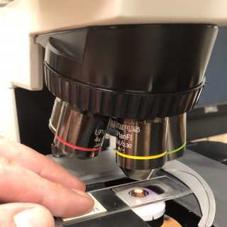
This podcast is part of the 2020 NSH Symposium/Convention Poster Podcast Series. Authors: Damien Laudier, Laudier Histology
Paul J. Wang: Welcome to the monthly podcast On the Beat for circulation, arrhythmia, and electrophysiology. I'm Dr. Paul Wang, Editor In Chief, with some of the key highlights from this month's issue. Elizabeth Wang and Associates examined the relationship between acute precipitants of atrial fibrillation and long-term recurrence of atrial fibrillation, AF, from a multi-institutional, longitudinal electronic medical record database. Among 10,723 patients with newly diagnosed Afib, age 67.9 years, 41% women, the authors found that 19% had an acute AF precipitant, the most common of which were cardiac surgery in 22%, pneumonia in 20% and non-cardiothoracic surgery in 15%. The cumulative incidence of AF recurrence at five years was 41% among individuals with a precipitant, compared to 52% in those without a precipitant. Adjusted hazard ratio 0.75 P < 0.001. The lowest risk of recurrence among those with precipitants with postoperative atrial fibrillation, five-year incidence 32% in cardiac surgery and 39% in non-cardiothoracic surgery. Regardless of the initial precipitant, recurrent atrial fibrillation was associated with an increased adjusted risk of heart failure, hazard ratio of 2.74 P < 0.001, Stroke, hazard ratio 1.57 P < 0.001 and mortality, hazard ratio 2.96 P < 0.001. Thus, the authors found that atrial fibrillation after acute precipitant frequently recurs and the recurrence is associated with substantial long-term morbidity and mortality. In the next paper, Jacob Koruth and associates examine the effect of pulse field ablation on the esophagus in a novel in-vivo porcine esophageal injury model. The authors studied 10 animals under general anesthesia while the lower esophagus was deflected towards the inferior vena cava using an esophageal deviation balloon and ablation was formed from within the inferior vena cava at areas of esophageal contact. Six animals received eight pulse field ablation applications per site and four animals received six clusters of irrigated radio frequency ablation applications at 30 Watts for 30 seconds. All animals survived to 25 days, sacrificed, and the esophagus was submitted for a pathological examination including 10 discreet histological sections of the esophagus. The authors found that zero out of six pulse field ablation animals demonstrated esophageal lesions while esophageal injury occurred in all four radio frequency ablation animals, P = 0.005. A mean of 1.5 mucosal lesions per animal, length 21.8 millimeters with 4.9 millimeters were observed, including one esophageal pulmonary fistula, and deep esophageal ulcers in the other animals. Histological examination demonstrated tissue necrosis surrounded by an acute and chronic inflammation and fibrosis. The necrotic radio frequency ablation lesions involved multiple esophageal tissue layers with evidence of arteriolar medial thickening and fibrosis of peri-esophageal nerves, abscess formation and full thickness esophageal wall disruption were seen in the areas of perforation or fistula. In our next paper, Peter Noseworthy and associates examine whether the ability of deep learning algorithms to detect low left ventricular ejection fraction using the 12 lead electrocardiogram varies by race or ethnicity. The authors used a retrospective cohort analysis and included 97,829 patients with paired electrocardiograms and echocardiograms and used a convolutional neural network to identify patients with a left ventricular ejection fraction less than or equal to 35% from the 12 lead electrocardiogram. The convolutional neural network was previously derived in a homogeneous population, 96.2% non Hispanic white, N = 44,959 which demonstrated consistent performance to detect low left ventricular ejection fraction across a range of racial ethnic subgroups in a separate cohort of 52,870 patients (Non-Hispanic white 44,524 patients with an AUC of 0.93; Asian 557 with an AUC of 0.96; Black/African American N = 651 with an AUC of 0.937; in Hispanic/Latino N = 331 AUC of 0.937; in Native American/Alaskan N = 223 AUC of 0.938). In secondary analysis, a separate neural network was able to discern racial subgroup category, Black/African American AUC 0.84 and white non-Hispanic AUC 0.75 in a five-class classifier. In a network trained only in non-Hispanic whites, from the original derivation cohort, performed similarly well across a range of racial ethnic subgroups in the testing cohort with at least an AUC of 0.93 in all racial ethnic subgroups. The authors concluded that while ECG characteristics vary by race, this did not impact the ability of a convolutional neural network to predict low left ventricular ejection fraction from the ECGs. They recommend reporting of performance against diverse ethnic, racial, age, and gender groups for all new artificial intelligent tools. In our next paper, Benjamin Shoemaker and associates examine the association between atrial fibrillation or AF genetic susceptibility and recurrence after de novo AF ablation, using a comprehensive polygenic risk score for AF in the 10 centers from the AF genetics consortium. AF genetic susceptibility was measured using a previously described a polygenic risk score, N = 929 snips. The overall arrhythmia recurrence rate between 3 and 12 months was 44% in 3,259 patients. Patients with a higher AF genetic susceptibility were younger and have fewer clinical risk factors for atrial fibrillation. Persistent atrial fibrillation has a ratio of 1.39, left atrial size has a ratio of 1.32, and left ventricular ejection fraction per 10% has a ratio of 0.88, were associated with increased risk of occurrence. In unit varied analysis, the authors found that AF genetic susceptibility had a hazard ratio of 1.08 P = 0.07 and in multivariate analysis hazard ratio 1.06 with a P value 0.13. In our next paper, Mohit Turagam and associates reported the outcomes of the first inhuman value trial, which uses low intensity collimated ultrasound or LICU guided anatomical mapping in robotic ablation to isolate the pulmonary veins for atrial fibrillation ablation. In 52 paroxysmal atrial fibrillation patients, ultrasound M-mode based left atrial anatomies were successfully created and ablation was performed under robotic control along an operated defined lesion path. The operatives found that acute pulmonary vein isolation was achieved in 98% of pulmonary veins using LICU only in 77% of pulmonary veins and requiring touch-up with a standard radio frequency ablation catheter in 23% of the pulmonary veins. The touch up rate decreased to 5.8% in patients undergoing LICU ablation with an enhanced software. Freedom from atrial relational recurrence was 79.6% at 12 months or 92.3%, 12 out of 13 patients with the enhanced software. Major adverse events occurred in three patients or 5.8%. One had transient diaphragmatic paralysis, one vascular access complication and one had transient ST segment elevation from air-embolism without sequelae. In our next paper, Miguel Rodrigo and associates mapped electrical patterns of disorganization and reasons of reentrant activity in atrial fibrillation, or AF, from the body surface using electrocardiographic imaging. The author examined the bi-atrial intracardiac electrograms of 47 patients at ablation (30 persistent, 29 males, age 63 years) obtained using 64-pole basket catheters while simultaneously recording 57-lead body surface electrocardiogram. The authors found the body surface mapping showed greater atrial fibrillation organization near intracardiac detected drivers and elsewhere, both in phase singularity density in numbers of drivers, they found that complexity defined as a number of stable AF reentrant sites was concordant between the noninvasive and invasive methods. The subset receiving targeted ablation, AF complexity, showed lower values in those in whom AF terminated than in those in whom AF did not terminate, P < 0.01. The authors concluded that AF complexity, assessed noninvasively, correlates well with organized, disorganized regions detected by intracardiac mapping. In our next paper, Krystien Lieve and Veronica Dusi and associates examined whether heart rate reduction immediately after exercise is regulated by autonomic reflexes, particularly vagal tone and may be associated with symptoms and ventricular arrhythmias in patients with catecholaminergic polymorphic ventricular tachycardia, CPVT. In a retrospective observational study, the authors studied 187 patients mean age 36 years, 68 or 36% symptomatic before diagnosis, pre-exercise stress test heart rate and maximal heart rate were equal amongst symptomatic and asymptomatic patients. Patients that were symptomatic prior to diagnosis had a greater delta HRR one prime after a maximum exercise, 43 versus 25, P < 0.001. Corrected for age, gender, and relatedness, patients in the upper tertile for Delta HRR one prime had an odd ratio of 3.4 of being symptomatic before diagnosis, P < 0.001. In addition, Delta HRR one prime was higher in patients with complex ventricular arrhythmias at exercise stress test, off antiarrhythmic drugs. After diagnosis, patients with a Delta HRR one prime in the upper tertile of its distribution, had significantly more rhythmic events as compared to patients and other tertiles, P=0.045. The authors concluded that CPVT patients with a larger heart rate reduction following exercise are more likely to be symptomatic and have complex ventricular arrhythmias during first exercise stress test off antiarrhythmic drugs. In our next paper, Balvinder Handa and associates examined whether low spatial resolution, sequentially acquired data can be used to examine the global fibrillation organization, characterizing dominant propagating patterns and identifying rotational drivers. The authors employed ranger causality analysis, an econometric tool for quantifying causal relationships between complex time series, which was developed as a novel fibrillation mapping tool and adapted to low spatial resolution sequentially acquired data. Ventricular fibrillation, or VF, optical mapping was performed and Langendorff-perfused Sprague Dawley rat hearts, N = 18. And novel algorithms were developed using Granger causality analysis to quantify causal dependence of neighboring signals and plot Granger causality analysis vectors, quantify global organization using causality pairing index, a measure of neighboring causal signal pairs, and localize rotational drivers by quantifying the circular interdependence of neighboring signals with the circular interdependence value. Granger causality analysis based mapping tools were optimized for low spatial resolution by down sampled optical mapping data validated against high resolution phase mapping analysis and further tested in previous VF optical mapping recordings of coronary perfused donor heart LV wedge preparations, N = 12, and adaptive for sequentially acquired intracardiac electric Grande during human persistent atrial relation mapping, N=16. The authors found that global VF organization quantified by causality pairing index showed a negative correlation at progressively lower resolutions in organized VF with high causality pairing index values. Ranger causality analysis vector mapping characterize dominant propagating patterns and localized stable rotational drivers with the circular interdependence value showing a significant difference in driver versus non driver regions, P = 0.0002. These findings were further confirmed in human VF in persistent atrial fibrillation, a positive correlation was found between causality peri-index and presence of stable rotational drivers. 50% of patients had rotational drivers with a low incidence of 0.9 rotational drivers per patient. In a special report, Piotr Futyma and associates report on the use of bipolar radiofrequency ablation of ventricular arrhythmias originating in the vicinity of the His bundle. Bryce Alexander and associates report in a research letter the patient acceptance of cybersecurity upgrade in ICDs. That's it for this month. We hope that you'll find the journal to be the go to place for everyone interested in the field. See you next time. This program is copyright American Heart Association 2020.

This mini episode covers erythema toxicum, AKA erythema toxicum neonatorum, toxic erythema of the newborn, and newborn rash. Erythema toxicum is a very common rash in newborns, especially term babies, and is benign and self resolving. In this episode, we discuss: Typical features of the newborn rash Differential diagnoses to consider How to describe newborn rash to parents Histological features of newborn rash Investigations that clinicians may consider if diagnosis if unclear Links and resources: Follow us on Instagram: https://www.instagram.com/yourekiddingright.pod/ and Facebook: https://www.facebook.com/yourekiddingrightpod-107273607638323/ Our email is yourekiddingrightpod@gmail.com Make sure you hit SUBSCRIBE/FOLLOW so you don’t miss out on any pearls of wisdom and RATE if you can to help other people find us! (This isn’t individual medical advice, please use your own clinical judgement and local guidelines when caring for your patients)
Link to bioRxiv paper: http://biorxiv.org/cgi/content/short/2020.11.05.369900v1?rss=1 Authors: Toro, C. A., Hansen, J., Siddiq, M. M., Johnson, K., Zhao, W., Azulai, D., Das, D. K., Bauman, W., Sebra, R., Cai, D., Iyengar, R., Cardozo, C. P. Abstract: Spinal cord injury (SCI) is a devastating form of neurotrauma. Patients who carry one or two ApoE4 alleles show worse functional outcomes and longer hospital stays after SCI but the cellular and molecular underpinnings for this genetic link remain poorly understood. Thus, there is a great need to generate animal models to accurately replicate the genetic determinants of outcomes after SCI to spur development of treatments that improve physical function. Here, we examined outcomes after a moderate contusion SCI of transgenic mice expressing human ApoE3 or ApoE4. ApoE4 mice have worse locomotor function and coordination after SCI. Histological examination revealed greater glial staining in ApoE4 mice after SCI associated with reduced levels of neuronal sprouting markers. Bulk RNA sequencing revealed that subcellular processes (SCPs), such as extracellular matrix organization and inflammatory responses, were highly-ranked among upregulated genes at 7 days after SCI in ApoE4 variants. Conversely, SCPs related to neuronal action potential and neuron projection development were increased in ApoE3 mice at 21 days. In summary, our results reveal a clinically relevant SCI mouse model that recapitulates the influence of ApoE genotypes on post-SCI function in individuals who carry these alleles and suggest that the mechanisms underlying worse recovery for ApoE4 animals involve glial activation and loss of sprouting and synaptic activity. Copy rights belong to original authors. Visit the link for more info
Link to bioRxiv paper: http://biorxiv.org/cgi/content/short/2020.10.21.347062v1?rss=1 Authors: Miguez, A., Fernandez-Garcia, S., Monguio-Tortajada, M., Bombau, G., Galofre, M., Garcia-Bravo, M., Vila, C., Sanders, P., Fernandez-Medina, H., Poquet, B., Salado-Manzano, C., Roura, S., Alberch, J., Segovia, J. C., Allen, N. D., Borras, F. E., Canals, J. M. Abstract: Research on neurodegenerative disorders has been hampered by the limited access to patients' brain tissue and the absence of relevant physiological models with human neurons, accounting for the little success of clinical trials. Moreover, post-mortem samples cannot provide a detailed picture of the complex pathological mechanisms taking place throughout the course of the disease. This holds particularly true for Huntington's disease (HD), an incurable inherited brain disorder marked by a massive striatal degeneration due to abnormal accumulation of misfolded huntingtin protein. To characterize progressive human neurodegeneration in vivo, we transplanted induced pluripotent stem cell-derived human neural progenitor cells (hNPCs) from control (CTR-hNPCs) and HD patients (HD-hNPCs) into the striatum of neonatal wild-type mice. Implanted human cells were examined by immunohistochemistry and electron microscopy, and chimeric mice were subjected to behavioral testing. Most grafted hNPCs differentiated into striatal neurons that sent axonal projections to their natural targets and established synaptic connections within the host basal ganglia circuitry. HD-hNPCs first showed developmental abnormalities characterized by an increased proliferation and accelerated medium spiny neuron (MSN) differentiation, mimicking the initial striatal hypertrophy of child mutant huntingtin (mHTT) carriers. HD human striatal neurons progressively developed mHTT oligomers and aggregates, which primarily targeted mitochondria, endoplasmic reticulum and nuclear membrane to cause structural alterations. Five months after transplantation, selective death of human MSNs and striatal degeneration altered mouse behavior, suggesting disease propagation to non-mutated host cells. Histological analysis and co-culture experiments revealed that HD-hNPCs secreted extracellular vesicles containing soluble mHTT oligomers, which were internalized by mouse striatal neurons triggering cell death. Finally, in vivo pharmacological inhibition of the exosomal secretory pathway through sphingosine-1 phosphate receptor functional antagonism, limited the spreading of apoptosis within the host striatum. Our findings cast new light on human neurodegeneration, unveiling cell and non-cell autonomous mechanisms that drive HD progression in patients. Copy rights belong to original authors. Visit the link for more info
Link to bioRxiv paper: http://biorxiv.org/cgi/content/short/2020.10.04.324335v1?rss=1 Authors: Budak, M., Grosh, K., Corfas, G., Zochowski, M., Booth, V. Abstract: Hidden hearing loss (HHL) is an auditory neuropathy characterized by normal hearing thresholds but reduced amplitude of the sound-evoked auditory nerve compound action potential (CAP). It has been proposed that in humans HHL leads to speech discrimination and intelligibility deficits, particularly in noisy environments. Animal models originally indicated that HHL can be caused by moderate noise exposures or aging, and that loss of inner hair cell (IHC) synapses could be its cause. A recent study provided evidence that transient loss of cochlear Schwann cells also causes permanent auditory deficits in mice which have characteristics of HHL. Histological analysis of the cochlea after auditory nerve remyelination showed a permanent disruption of the myelination patterns at the heminode of type I spiral ganglion neuron (SGN) peripheral terminals, suggesting that this defect could be contributing to HHL. To shed light on the mechanisms of different HHL scenarios and to test their impact on type I SGN activity, we constructed a reduced biophysical model for a population of SGN peripheral axons. We found that the amplitudes of simulated sound-evoked SGN CAPs are lower and have greater latencies when the heminodes are disorganized, i.e. they are placed at different distances from the hair cell rather than at the same distance as seen in the normal cochlea. Thus, our model confirms that disruption of the position of the heminode causes desynchronization of SGN spikes leading to a loss of temporal resolution and reduction of the sound-evoked SGN CAP. We also simulated synaptopathy by removing high threshold IHC-SGN synapses and found that the amplitude of simulated sound-evoked SGN CAPs decreases while latencies remain unchanged, corresponding to what has been observed in noise exposed animals. This model can be used to further study the effects of synaptopathy or demyelination on auditory function. Copy rights belong to original authors. Visit the link for more info

Histological specimens are recommended to assess a patient's PD-L1 status; however, it is not always easy or even feasible to secure a histological specimen in advanced cancers such as stage IV NSCLC. Cytological specimens are typically used in this setting to assess other molecular markers such as ALK or ROS1 – should they also be used to assess PD-L1 status? This episode summarises the evidence for and against a cytological approach, before joining Professor Frederique Penault-Llorca for her opinion on whether cytology should be used to assess PD-L1 status in real-world practice. References: Gosney J, et al. Lung Cancer. 2020; 141:101-106 Heymann J, et al. Cancer Cytopathology. 2017; 125(12): 896-907 Wagner C, et al. Eur Respir J. 2018; 52: Suppl. 62, PA2217. Access more free education today! Visit the website, follow us on Twitter (@onckip) or connect on LinkedIn. This independent educational activity is supported by an educational grant from Merck, Sharpe & Dohme Corp. The educational content has been developed by Liberum IME in conjunction with an independent steering committee; MSD corp. has had no influence on the content of this education.
Link to bioRxiv paper: http://biorxiv.org/cgi/content/short/2020.09.09.289025v1?rss=1 Authors: Telegin, G. B., Minakov, A. N., Chernov, A. S., Kazakov, V. A., Kalabina, E. A., Belogurov, A. A., Konovalov, N. A., Gabibov, A. G. Abstract: Up to 500,000 people worldwide suffer from spinal cord injuries (SCI) annually, according to the WHO. Animal models are essential for searching novel methodological guidelines and therapeutic agents for SCI treatment. We developed an original model of posttraumatic spinal cord glial scar in rats using cryoapplication. The method is based on cryodestruction of spinal cord tissue with liquid nitrogen. Thirty six male SD linear rats of SPF category were included in this experimental study. A T13 unilateral hemilaminectomy was performed with an operating microscope, as it was extremely important not to penetrate the dura mater, and liquid nitrogen was applied into the bone defect for one minute. The animals were euthanized at various intervals ranging from 1 to 60 days after inducing cryogenic trauma, their Th12-L1 vertebrae were removed "en bloc" and the segment of the spinal cord exposed to the cryoapplicator was carefully separated for histological examination. The study results demonstrated that cryoapplication of liquid nitrogen, provoking a local temperature of approximately minus 20 degrees Celsius, produced a highly standardized transmural defect which extended throughout the dorsoventral arrangement of the spinal cord and had an "hour-glass" shape. During the entire study period (1-60 post-injury days), the glial scarring process and the spinal cord defect were located within the surgically approached vertebral space (Th13). Unlike other available experimental models of SCI (compression, contusion, chemical, etc.), the present option is characterized by a minimal invasiveness (the hemilaminectomy is less than 1 mm wide), high precision and consistency. Also, there was a low interanimal variability in histological lesions and dimensions of the produced defect. The original design of cryoapplicator used in the study played a major role in achieving these results. The original technique of high-precision cryoapplication for inducing consistent morphodynamic glial scarring could facilitate a better understanding of the self-recovery processes of injured spinal cord and would be helpful for proposing new platforms for the development of therapeutic strategies. Copy rights belong to original authors. Visit the link for more info

HOPA Now is the official podcast of the Hematology/Oncology/Pharmacy Association, an organization dedicated to supporting pharmacy practitioners and promoting the advancement of Hematology/Oncology/Pharmacy to optimize the care of individuals impacted by cancer. These educational podcasts are part of our BCOP Preparatory and Recertification Course, which is designed to prepare oncology pharmacists preparing to sit for the BCOP Certification Exam, as well as meet the BPS requirement to complete a BCOP Preparatory/Recertification Review Course. In this episode of HOPA Now, Dr. Brandon Shank offers an overview of the fundamentals of lymphomas, including biopsy recommendations, histological determinations, and screenings that are commonly administered during treatment. He highlights the indications for starting follicular lymphoma treatment and shares guidelines and criteria for administering high dose methotrexate, and concludes with information regarding various B-cell lymphomas and mycosis fungoides. In this episode you will learn: Lymphomas Fundamentals Biopsy recommendations for making an accurate diagnosis Histological determination through immunotyping used to determine subclassifications Common screenings that are administered during treatment Understanding Rituximab and the biosimilars and NCCN recommendations Indications for starting follicular lymphoma treatment Guidelines and criteria for administering high dose methotrexate and dose-adjusted R-EPOCH Prevention of bacterial and other infections Risk factors and recommendations associated with various B-cell lymphomas and mycosis fungoides Mentioned in This Episode: HOPA Tweetables: “In select patients who have a high hepatitis B antibody level, they may not need prophylaxis.” — Dr. Brandon Shank “There are important things to consider to make sure you’re monitoring patients that are receiving high-dose methotrexate appropriately.” — Dr. Brandon Shank “Dose-adjusted R-EPOCH is a special regiment where the dose is adjusted based on the patient's specific factors.” — Dr. Brandon Shank

Cancer Grand Rounds Lectures from the Norris Cotton Cancer Center Podcasts
Norris Cotton Cancer Center Grand Rounds February 4, 2020 Saeed Hassanpour, PhD Associate Professor Biomedical Data Science Associate Professor of Epidemiology

This year, Halloween falls on a Thursday. So we've put together a special episode to celebrate it! But first...a question: What do Frankenstein's monster, Spock, and Rick Deckard all have in common? Find out in this week's episode of BrainWaves. Produced by James E. Siegler. Special guest, Scott Kasner (Hospital of the University of Pennsylvania) with fair use of material from Young Frankenstein (1974), Star Trek: The Original Series (1966-1969), and Blade Runner (1982). Music courtesy of Grossman, Ewell and Grainger, Heftone Banjo Orchestra, Sergey Chereminisov, and Ondrosik under a Creative Commons License. Sound effects by Mike Koenig. BrainWaves' podcasts and online content are intended for medical education only and should not be used for clinical decision making. Be sure to follow us on Twitter @brainwavesaudio for the latest updates to the podcast. REFERENCES Labbe R, Firl A, Jr., Mufson EJ and Stein DG. Fetal brain transplant: reduction of cognitive deficits in rats with frontal cortex lesions. Science. 1983;221:470-2. Brain transplant for Parkinson's disease. Lancet. 1987;1:1012-3. Ignacio V, Collins VP, Suard IM and Jacque CM. Survival of astroglial cell lineage from adult brain transplant. Dev Neurosci. 1989;11:175-8. Landau WM. Clinical neuromythology VII. Artificial intelligence: the brain transplant cure for parkinsonism. Neurology. 1990;40:733-40. Freed CR, Breeze RE, Rosenberg NL, Schneck SA, Kriek E, Qi JX, Lone T, Zhang YB, Snyder JA, Wells TH and et al. Survival of implanted fetal dopamine cells and neurologic improvement 12 to 46 months after transplantation for Parkinson's disease. The New England journal of medicine. 1992;327:1549-55. Landau WM. Clinical neuromythology X. Faithful fashion: survival status of the brain transplant cure for parkinsonism. Neurology. 1993;43:644-9. Grabowski M, Johansson BB and Brundin P. Neocortical grafts placed in the infarcted brain of adult rats: few or no efferent fibers grow from transplant to host. Exp Neurol. 1995;134:273-6. Schulz MK, McNulty JA, Handa RJ, Hogan TP, Tillotson GL, Shaw PL, Zimmer J and Castro AJ. Fetal neocortical transplants grafted into neocortical lesion cavities made in newborn rats: an analysis of transplant integration with the host brain. Cell Transplant. 1995;4:123-32. Deacon T, Schumacher J, Dinsmore J, Thomas C, Palmer P, Kott S, Edge A, Penney D, Kassissieh S, Dempsey P and Isacson O. Histological evidence of fetal pig neural cell survival after transplantation into a patient with Parkinson's disease. Nat Med. 1997;3:350-3. Freed CR, Greene PE, Breeze RE, Tsai WY, DuMouchel W, Kao R, Dillon S, Winfield H, Culver S, Trojanowski JQ, Eidelberg D and Fahn S. Transplantation of embryonic dopamine neurons for severe Parkinson's disease. The New England journal of medicine. 2001;344:710-9. Bhattacharya N, Chhetri MK, Mukherjee KL, Ghosh AB, Samanta BK, Mitra R, Bhattacharya M, Bhattacharya S and Bandopadhyaya T. Can human fetal cortical brain tissue transplant (up to 20 weeks) sustain its metabolic and oxygen requirements in a heterotopic site outside the brain? A study of 12 volunteers with Parkinson's disease. Clin Exp Obstet Gynecol. 2002;29:259-66. Kumar R and Mahapatra AK. Concept of brain transplant in pre-historic era. Childs Nerv Syst. 2009;25:393-4. Bjorklund A, Dunnett SB, Stenevi U, Lewis ME and Iversen SD. Reinnervation of the denervated striatum by substantia nigra transplants: functional consequences as revealed by pharmacological and sensorimotor testing. Brain Res. 1980;199:307-33.

In this podcast Imogen Johns discusses histological evaluation of liver biopsies, and Sam Hole discusses equine odontoclastic tooth resorption and hypercementosis. https://onlinelibrary.wiley.com/doi/10.1111/eve.12871 https://onlinelibrary.wiley.com/doi/10.1111/eve.12603
This podcast is part of the 2017 NSH Symposium/Convention Poster Podcast Series. The lead author of this poster is Ruth Fidler. For more information on the author and to view the abstract, visit The Block.

The purpose of this study was to perform a histological comparative analysis of tibiotalar joint samples taken from areas of osteolysis adjacent to total ankle arthroplasties vs control synovial specimens to determine the reaction to and presence of polyethylene (PE) particles. This study is the largest ankle arthroplasty histological analysis to show that areas of osteolysis consist of abundant polyethylene wear particles, present both intracellularly and extracellularly. Furthermore, these areas were associated with a CD163+ macrophage infiltrate and frequently a foreign body reaction with giant cells engulfing PE particles. It is likely that implant wear particles play a significant role in osteolysis based on the histopathology. To view the article, click here.

Background: Oronasal fistulas are a frequent complication after cleft palate surgery. Numerous repair methods have been described, but wound-healing problems occur often. The authors investigated, for the first time, the suitability of multilayered amniotic membrane allograft for fistula repair in a laboratory experiment (part A), a swine model (part B), and an initial patient series (part C). Methods: In part A, one-, two-, and four-layer porcine and human amniotic membranes (n = 20 each) were fixed in a digital towing device and the force needed for rupture was determined. In part B, iatrogenic oronasal fistulas in 18 piglets were repaired with amniotic membrane allograft, autofetal amniotic membrane, or small intestinal submucosa (n = 6 each). Healing was evaluated by probing and visual inflammation control (no/moderate/strong) on postoperative days 3, 7, 10, and 76. Histological analysis was performed to visualize tissue architecture. In part C, four patients (two women and two men, ages 21 to 51 years) were treated with multilayered amniotic membrane allograft. Results: In part A, forces needed for amniotic membrane rupture increased with additional layers (p < 0.001). Human amniotic membrane was stronger than porcine membrane (p < 0.001). In part B, fistula closure succeeded in all animals treated with amniotic membrane with less inflammation than in the small intestinal submucosa group. One fistula remained persistent in the small intestinal submucosa group. In part C, all fistulas healed completely without inflammation. Conclusions: Amniotic membrane is an easily available biomaterial and can be used successfully for oronasal fistula repair. The multilayer technique and protective plates should be utilized to prevent membrane ruptures.
Dr. Louise Rodino-Klapac Discusses Alpha 7 Integrin As A Therapeutic Approach to Muscular Dystrophy Guest: Louise Rodino-Klapac, PhD, principal investigator, Center for Gene Therapy, The Research Institute at Nationwide Children’s Hospital. Access an abstract of this month’s featured research article: AAV-mediated Overexpression of Human a7 Integrin Leads to Histological and Functional Improvement in Dystrophic Mice. Mol Ther. 2013 Mar;21(3):520-5. doi: 10.1038/mt.2012.281. Epub 2013 Jan 15.
Dr. Louise Rodino-Klapac Discusses Alpha 7 Integrin As A Therapeutic Approach to Muscular Dystrophy Guest: Louise Rodino-Klapac, PhD, principal investigator, Center for Gene Therapy, The Research Institute at Nationwide Children’s Hospital. Access an abstract of this month’s featured research article: AAV-mediated Overexpression of Human a7 Integrin Leads to Histological and Functional Improvement in Dystrophic Mice. Mol Ther. 2013 Mar;21(3):520-5. doi: 10.1038/mt.2012.281. Epub 2013 Jan 15.

Although many aspects of ovarian differentiation have been established,comparatively little is known about prenatal follicle formation anddifferentiation of bovine ovaries. The objective of this investigationwas to study the role of the surface epithelium during the developmentof germ cell nests, germ cell cords and follicle formation in the fetalbovine ovary. Associated important proliferation and apoptotic featureswere further investigated. Additionally, the expression pattern of theS100 protein was detected. A strong increase of mitotic figures wasdetected in the surface epithelium, germ cell nests and germ cell cordsof ovaries with a crown-rump length (CRL) of 13.0-58.0 cm. Oocytes werepositively stained with S100 in bovine ovaries from fetuses with a CRLof 21.0 cm. The staining intensity enhanced parallel to increasingoocyte and follicle sizes during the ovary development. In later stages,a strong staining for S100 was observed in healthy oocytes incontradistinction to atretic oocytes where no expression of the S100protein could be found. In conclusion, increasing mitosis index ofsurface epithelium cells, as well as oogonia directly beneath thesurface epithelium, in combination with open surface connection duringstages from a CRL of 11.0-94.0 cm of bovine fetal ovaries could play animportant role in the period of time of ongoing folliculogenesis andderivation of granulosa cells. Additionally, S100-positive oocytes inprimordial and later follicle stages joined by a high rate ofKi67-positive index in surrounding granulosa cells indicate that in theoocytes the S100 protein can perhaps be a useful marker for intactoocytes in bovine ovaries.

Prof Christos Sotiriou discusses the results of the BIG-1-98 study. This trial demonstrated that the genomic grade index can separate histological grade 2 breast tumours into low or high categories with different clinical outcomes. Prof Sotiriou explains how this system could have considerable benefits to patients, reducing the level of chemotherapy required to treat low grade patients.

4th EUTROC (European Network for Translational Research in Ovarian Cancer
Dr Jozien Helleman talks to ecancer at the 4th EUTROC meeting in Liverpool, UK about a study that extensively categorised ovarian tumours. Methods used included sequencing and gene expression, as well as combining this with therapy response. Dr Helleman also discusses the potential for identifying drugable targets through these methods.

Dr. James Lee discusses his manuscript A Novel Histologic Scoring System to Evaluate Mucosal Biopsies From Patients With Eosinophilic Esophagitis. To view the print version of this abstract go to http://tinyurl.com/lbnkmd

Fri, 1 Jul 2011 12:00:00 +0100 http://gut.bmj.com/content/60/7/881.long https://epub.ub.uni-muenchen.de/21910/1/oa_21910.pdf Lamerz, Rolf
Background Although histopathological grading systems for disc degeneration are frequently used in research, they are not yet integrated into daily care routine pathology of surgical samples. Therefore, data on histopathological changes in surgically excised disc material and their correlation to clinical parameters such as age, gender or body mass index (BMI) is limited to date. The current study was designed to correlate major physico-clinical parameters from a population of orthopaedic spine center patients (gender, age and BMI) with a quantitative histologic degeneration score (HDS). Methods Excised lumbar disc material from 854 patients (529 men/325 women/mean age 56 (15-96) yrs.) was graded based on a previously validated histologic degeneration score (HDS) in a cohort of surgical disc samples that had been obtained for the treatment of either disc herniation or discogenic back pain. Cases with obvious inflammation, tumor formation or congenital disc pathology were excluded. The degree of histological changes was correlated with sex, age and BMI. Results The HDS (0-15 points) showed significantly higher values in the nucleus pulposus (NP) than in the annulus fibrosus (AF) (Mean: NP 11.45/AF 7.87), with a significantly higher frequency of histomorphological alterations in men in comparison to women. Furthermore, the HDS revealed a positive significant correlation between the BMI and the extent of histological changes. No statistical age relation of the degenerative lesions was seen. Conclusions This study demonstrated that histological disc alterations in surgical specimens can be graded in a reliable manner based on a quantitative histologic degeneration score (HDS). Increased BMI was identified as a positive risk factor for the development of symptomatic, clinically significant disc degeneration.

Background The proinflammatory cytokines interleukin 1 beta (IL-1 beta) and IL-18 are central players in the pathogenesis of inflammatory bowel disease (IBD). In response to a variety of microbial components and crystalline substances, both cytokines are processed via the caspase-1-activating multiprotein complex, the NLRP3 inflammasome. Here, the role of the NLRP3 inflammasome in experimental colitis induced by dextran sodium sulfate (DSS) was examined. Methods IL-1b production in response to DSS was studied in macrophages of wild-type, caspase-1(-/-), NLRP3(-/-), ASC(-/-), cathepsin B(-/-) or cathepsin L(-/-) mice. Colitis was induced in C57BL/6 and NLRP3(-/-) mice by oral DSS administration. A clinical disease activity score was evaluated daily. Histological colitis severity and expression of cytokines were determined in colonic tissue. Results Macrophages incubated with DSS in vitro secreted high levels of IL-1b in a caspase-1-dependent manner. IL-1b secretion was abrogated in macrophages lacking NLRP3, ASC or caspase-1, indicating that DSS activates caspase-1 via the NLRP3 inflammasome. Moreover, IL-1b secretion was dependent on phagocytosis, lysosomal maturation, cathepsin B and L, and reactive oxygen species (ROS). After oral administration of DSS, NLRP3(-/-) mice developed a less severe colitis than wild-type mice and produced lower levels of proinflammatory cytokines in colonic tissue. Pharmacological inhibition of caspase-1 with pralnacasan achieved a level of mucosal protection comparable with NLRP3 deficiency. Conclusions The NLRP3 inflammasome was identified as a critical mechanism of intestinal inflammation in the DSS colitis model. The NLRP3 inflammasome may serve as a potential target for the development of novel therapeutics for patients with IBD.

Tierärztliche Fakultät - Digitale Hochschulschriften der LMU - Teil 04/07
Histological investigation of the dorsal carpal joint integument of differently housed fattening bulls Aim of this trial was to investigate - both macro- and microscopically - the effects of various surfaces on the integument of dorsal carpal joints, the main stress zones when cattle rest and rise, using differently housed fattening bulls as samples. The histological differences, which result from the varying housing types, were described to assess possible postural deformities and their subsequent consequences for animal protection with the help of the respective re-sults. Additionally the functional circle “rest behaviour” with its respective motion sequence, which is important to the question at hand, was also included in the trial. Fattening bulls are usually held on concrete slatted floor, which serves as surface for moving and resting. Postural integument lesions at the joint regions exposed during the resting and rising process were previously not of scientific interest. They do, however, have a high rele-vance, not only for the assessment of the animals’ well-being, but also regarding possible sub-sequent clinical illnesses and their economic consequences. In this trial animals were divided in four groups (concrete slatted floor with 2,5-3 m2 = con-crete group, 32 animals; rubber topped slatted floor with 2,5 - 3.0 m2/animal = G1-group, 40 animals; rubber topped slatted floor with 4,5 - 5.0 m2/animal = G2-group, 40 animals; straw housing, 6 animals = straw group). The resting, rising and lie down behaviour of the animals was documented (only slatted floor groups) and the integument in the dorsal carpal joint area was macroscopically examined. Samples of the respective integument areas of each carpal joint were histologically examined after slaughtering. In addition samples were laterally taken from the animals housed on straw at the carpal joint integument. These were examined and analyzed as reference specimen for non-pressure-exposed integument (lateral control). The analysis of the resting, rising and lie down behaviour showed that the position change and in particular the stress on the carpal joints is delayed or avoided by animals using atypical motion patterns on concrete slatted floor. The macroscopic inspection of the integument within the area of the dorsal carpal joints con-cerning the trial parameters scab and swelling of connective tissue showed significantly in-creased values in the concrete group in comparison to G1 and G2. The carpal joints of the straw group could not be examined at the relevant time of trial due to high defilement. The histological investigation of the respective pressure-exposed integument areas showed a thickness increase in all experimental groups compared to the results of the lateral control group not exposed to pressure. All integument layers were affected by this increase in thick-ness, its effects increased from the straw over the rubber to the concrete group, while no dif-ferences could be accounted for between the two rubber groups. The so called bursae subcutis occurred in subcutaneous tissue areas with the slatted floor groups, which is to be regarded as adaption to extreme chronic pressure. As reaction to the increased epidermal proliferation rate an accordingly higher developed stratum papillare, which had and increased amount of elastic fibres in the dermis, was found in the concrete group, probably due to the increased shearing forces of the concrete slatted floor. Within the epidermis were the largest morphometric dif-ferences, to which the stratum corneum contributed the most in terms of numbers. Here the results of the concrete group displayed extreme values, which were about 8-times increased in comparison to the straw group. For the three slatted floor types the mitosis rates and the frequency of occurring paraceratosis hyperplasia (nucleated cells in the stratum corneum) correlated as signs of accelerated horni-fication with the layer thickness growth of the epidermis. Deviating values were documented in the straw group. An accumulated presence of inflammation cells could be observed in the concrete and the straw group; it was, however, not statistically significant. While the histological results sug-gested mechanically caused integument lesions as an inflammation cause in the concrete group, various findings pointed to a damage of the integrity of the epidermal barrier function, due to the chemical-physical influence of a wet, liquid manure-impregnated lie down area in the final weeks of life with the straw group. Various histochemical colourings were prepared for the assessment of the keratinisation and hornification of the epidermis. Significant differences between the investigated groups could, however, not be determined. The implemented investigations produced a comprehensive overview of the development of the dorsal carpal joint’s integument under the effects of various housing types and led to a histological explanation of macroscopically visible integument changes. The determined in-tegument changes at the dorsal carpal joint are to be regarded as an attempt of the integument to form a protection against increased mechanical stress. Nevertheless, in case of animals housed on concrete slatted floor, it does lead to the conclusion that a motion pattern which is covering demand and avoids damage is not possible with the rising and lying down process. This demonstrates the high animal-protection risk of this housing type.
Objective: The aim of the current prospective study was to analyse the validity of MRI based diagnosis of brainstem gliomas which was verified by stereotactic biopsy and follow-up evaluation as well as to assess prognostic factors and risk profile. Methods: Between 1998 and 2007, all consecutive adult patients with radiologically suspected brainstem glioma were included. The MRI based diagnosis of the lesions was made independently by an experienced neuroradiologist. Histopathological evaluation was performed in all patients from paraffin embedded specimens obtained by multimodal image guided stereotactic serial biopsy technique. Histopathological results were compared with prior radiological assessment. Length of survival was estimated with the Kaplan–Meier method and prognostic factors were calculated using the Cox model. Results: 46 adult patients were included. Histological evaluation revealed pilocytic astrocytoma (n=2), WHO grade II glioma (n=14), malignant glioma (n=12), metastasis (n=7), lymphoma (n=5), cavernoma (n=1), inflammatory disease (n=2) or no tumour/ gliosis (n=3). Perioperative morbidity was 2.5% (n=1). There was no permanent morbidity and no mortality. All patients with ‘‘no tumour’’ or ‘‘inflammatory disease’’ survived. Patients with low grade glioma and malignant glioma showed a 1 year survival rate of 75% and 25%, respectively; the 1 year survival rate for patients with lymphoma or metastasis was 30%. In the subgroup with a verified brainstem glioma, negative predictors for length of survival were higher tumour grade (p=0.002) and Karnofsky performance score (70 (p=0.004). Conclusion: Intra-axial brainstem lesions with a radiological pattern of glioma represent a very heterogeneous tumour group with completely different outcomes. Radiological features alone are not reliable for diagnostic classification. Stereotactic biopsy is a safe method to obtain a valid tissue diagnosis, which is indispensible for treatment decision.
The chance of an intrathyroidal occurrence of a parathyroid gland is about 1-3%. Among the causes of hyperparathyroidism, parathyroid cases occur in less than 1% of patients. Here we present the case of a 63 year old Saudi female suffering from an intrathryroidal parathyroid carcinoma. The suspicion coming from the clinical investigations that the removed tumor tissue may be a parathyroid carcinoma could be confirmed by histology. Additionally non-radioactive in situ hybridization to localize mRNA transcripts for Cyclin D1 and immunohistochemical localization of Cyclin D1 was performed. Although parathyroid adenoma and carcinoma have disparate natural history, it can be difficult to differentiate between the two entities. Clinical presentation, operative findings may raise suspicion, but may not be conclusive especially if there is no evidence of invasion or metastasis, especially if the gland was intrathyroidal.
Objective: Liver regeneration is mainly based on cellular self-renewal including progenitor cells. Efforts have been made to harness this potential for cell transplantation, but shortage of hepatocytes and premature differentiated progenitor cells from extra-hepatic organs are limiting factors. Histological studies implied that resident cells in adult liver can proliferate, have bipotential character and may be a suitable source for cell transplantation. Methods: Particular cell populations were isolated after adequate tissue dissociation. Single cell suspensions were purified by Thy-1 positivity selection, characterised in vitro and transplanted in immunodeficient Pfp/Rag2 mice. Results: Thy-1+ cells that are mainly found in the portal tract and the surrounding parenchyma, were isolated from surgical liver tissue with high yields from specimens with histological signs of regeneration. Thy-1+ cell populations were positive for progenitor (CD34, c-kit, CK14, M2PK, OV6), biliary (CK19) and hepatic (HepPar1) markers revealing their progenitor as well as hepatic and biliary nature. The potential of Thy-1+ cells for differentiation in vitro was demonstrated by increased mRNA and protein expression for hepatic (CK18, HepPar1) and biliary (CK7) markers during culture while progenitor markers CK14, chromogranin A and nestin were reduced. After transplantation of Thy-1+ cells into livers of immunodeficient mice, engraftment was predominantly seen in the periportal portion of the liver lobule. Analysis of in situ material revealed that transplanted cells express human hepatic markers HepPar1 and albumin, indicating functional engraftment. Conclusion: Bipotential progenitor cells from human adult livers can be isolated using Thy-1 and might be a potential candidate for cell treatment in liver diseases.
Background: Whereas various molecular working mechanisms of shock waves have been demonstrated, no study has assessed in detail the influence of varying energy flux densities (EFD) on new bone formation in vivo. Methods: Thirty Chinchilla bastard rabbits were randomly assigned to 5 groups (EFD 0.0, 0.35, 0.5, 0.9 and 1.2 mJ/mm(2)) and treated with extracorporeal shock waves at the distal femoral region (1,500 pulses; 1 Hz frequency). To investigate new bone formation, animals were injected with oxytetracycline at days 5-9 after shock wave application and sacrificed on day 10. Histological sections of all animals were examined using broad-band epifluorescent illumination, contact microradiography and Giemsa-Eosin staining. Results: Application of shock waves induced new bone formation beginning with 0.5 mJ/mm(2) EFD and increasing with 0.9 mJ/mm(2) and 1.2 mJ/mm(2). The latter EFD resulted in new bone formation also on the dorsal cortical bone; cortical fractures and periosteal detachment also occurred. Conclusion: Here, for the first time, a threshold level is presented for new bone formation after applying shock waves to intact bone in vivo. The findings of this study are of considerable significance for preventing unwanted side effects in new approaches in the clinical application of shock waves. Copyright (c) 2008 S. Karger AG, Basel.

Fakultät für Biologie - Digitale Hochschulschriften der LMU - Teil 02/06
Multiple sclerosis (MS) is an autoimmune inflammatory disease of the central nervous system (CNS). Myelin antigen(s) specific T cells, B cells, and antibodies are thought to play a role in the pathogenesis of MS. While the influence of autoantigenspecific CD4+ T cells has been extensively studied in animal models, the relevance of autoantigen specific B cells and their interactions with pathogenic T cells are largely unknown. The original aim of the present study was to create a new mouse model with which to investigate the interaction of myelin autoantigen specific B and T cells and their role in MS pathogenesis. The study was further expanded to analyze the nature and triggers of spontaneous disease and similarity of the mouse lesion pattern to that in human disease. The double-transgenic mouse (“Devic mouse”) strain presented here contains myelin oligodendrocyte glycoprotein (MOG)-specific T as well as B cells. A significant proportion (>50%) of these mice showed spontaneous experimentalautoimmune encephalomyelitis (EAE)-like disease at a young age. In contrast, all single transgenic littermates were free of clinical disease. Spontaneous EAE requires both MOG-specific T and B cells, since the breeding of MOG-specific Ig heavy chain knock-in mice with ovalbumin specific T cell receptor (TCR) transgenic mice did not develop any disease. Histological analysis of the CNS of affected mice revealed restricted localization of lesions in the spinal cord and optic nerves as well as severe demyelination and axonal damage that spared brain and cerebellum. The inflammatory infiltrates were predominantly composed of macrophages and CD4+ T cells, but occasionally also eosinophils. This peculiar localization of the demyelinating lesions and infiltration profile differ from classic EAE and is reminiscent of Devic’s neuromyelitis optica, a variant of classic MS in humans. It is not well understood what triggers the initiation of spontaneous EAE. The microbial environment does not significantly affect the clinical disease. Stimulation of the innate immune system with toll-like receptor (TLR) ligands or depletion of putative regulatory cells did not significantly affect EAE development. The (re-)activation of lymphocytes in sick Devic mice mainly occurs in the CNS without evidence of priming in the peripheral lymphoid organs. MOG-specific B and T cells cooperate by means of several mechanisms. MOGspecific B cells, which bind MOG but not the immunodominant peptide MOG 35-55 via their surface immunoglobulin (Ig), efficiently presented even high dilutions of MOG to T cells. This resulted in the enhanced proliferation of T and B cells as well as rapid activation. Stimulated T, but not B cells, secreted large amounts of Th1 cytokines IFNg and IL-2 along with small amounts of Th2 cytokine IL-5. In addition, MOG-stimulated T and B cells expressed a set of co-stimulatory molecules, which further help to modulate the proliferation and activation. Surprisingly, the doubletransgenic Devic mice, but not their single transgenic littermates, had high titers of MOG-specific IgG1 antibodies in the serum, which indicates a previous encounter with antigen in vivo. However, similar MOG-specific serum IgG1 titers were present irrespective of the clinical status. The transfer of EAE by Devic splenocytes in immunodeficient mice or by bone marrow reconstitution in wild-type mice further supported the in vivo cooperation of MOG-specific T and B cells to induce spontaneous EAE. In summary, Devic mice show several salient features that are important for study of the pathogenic mechanisms of CNS autoimmunity. As a model of spontaneous autoimmunity, they may allow us to study the triggering factors of autoimmunity as well as the factors that determine restricted infiltration of immune cells into the CNS.In addition, the model may be useful for validating novel therapies for autoimmune CNS diseases.

Objective: To evaluate the staining characteristics of bromphenol blue used during vitreoretinal surgery in humans.Patients and methods: 13 patients with epiretinal membranes were included. Before and after surgery a complete clinical examination including best corrected visual acuity, funduscopy, fluorescein angiography, OCT (Stratus), Goldmann perimetry and multifocal ERG as well as photography of the macular area was performed. Bromphenol blue was used in concentrations of 0.2% in most patients. Removed epiretinal tissue was evaluated using electron microscopy.Results: Using dye concentrations of 0.2% a good demarcation of epiretinal membranes was seen in 11/13 patients. Staining of vitreous remnants at the vitreous base was seen in all patients. No dye-related adverse events were seen during follow-up in the functional tests (VA, ERG, perimetry) performed. Histological evaluation of epiretinal membranes showed unremarkable aspects of epiretinal cellular layers and unremarkable retinal surface of the internal limiting membrane (ILM).Conclusion: Bromphenol blue appears to be a very helpful and safe tool in posterior segment surgery. The staining characteristics need to be further evaluated in prospective study settings and larger numbers of patients.

Tierärztliche Fakultät - Digitale Hochschulschriften der LMU - Teil 02/07
Histological, immunhistochemical and molecular biological characterisation of retrovirus-induced murine haematopoietic neoplasms The murine leukaemia virus Akv has lymphomagenic properties in newborn inbred NMRI mice. The enhancer in the U3 long terminal repeat sequence of the Akv genome is a major determinant of retroviral lymphomagenesis. In this study, the pathogenicity of wild-type Akv, Akv1-99, an Akv lacking one 99 bp repeat in the U3 sequence, and a panel of ten Akv1-99 mutants containing mutations in the different transcription factor binding sites, was analysed. The retrovirus-induced haematopoietic tumours were classified according to the Bethesda Proposals (94, 138), based on histological and immunohistochemical analyses, and IgH gene rearrangements. All viruses were pathogenic. Tumour incidence ranged from 90 % to 100 %. The mutant viruses, however, differed in their latency periods and tissue specificity. Akv induced diffuse large B-cell lymphomas (32.5 %), follicular B-cell lymphomas (22.5 %), splenic marginal zone lymphomas (5 %), small B-cell lymphomas (5 %), and plasmacytomas (35 %). The mutant viruses induced almost exclusively plasmacytomas (up to 100 %). The latency periods of three mutant viruses were comparable to that of Akv; six mutant viruses showed a shorter latency (five latency periods were significantly shorter), and two mutant viruses with an additional mutation in the EGRE binding site induced tumours after significantly prolonged latency. These results suggest that mutations of transcription factor binding sites in the Akv enhancer maintained the pathogenicity of the viruses but resulted in a smaller spectrum of haematopoietic tumours with different impact on the latency. Comprehensive histological, immunohistochemical, and molecular analyses of 323 retrovirus-induced haematopoietic tumours (83.8 % plasmacytomas, 6.9 % diffuse large B-cell lymphomas, 4.5 % follicular B-cell lymphomas, 4.2 % splenic marginal zone lymphomas, 1.2 % small B-cell lymphomas, 1.2 % acute myeloid leukaemias, and 0.9 % precursor-T-cell lymphoblastic lymphomas) illustrated the importance of transcription factor binding sites of Akv and its impact on viral virulence and disease specificity. This model facilitates the dissection of disease-determining sequences in the retroviral genome and discloses differences and similarities between human and murine haematopoietic neoplasms.
Because of different measurement techniques and the easier design of the CRM prototype, this in vitro study aimed to compare the diagnostic performance and reproducibility of two electrical methods (Electronic Caries Monitor III, ECM and Cariometer 800, CRM) for occlusal caries detection, and to evaluate the effect of staining/ discoloration of fissures on diagnostic performance. Hundred and seventeen third molars with no apparent occlusal cavitation were selected. Six examiners inspected all specimens independently, using the CRM, and a subgroup of 4 using the ECM. Histological validation using a stereomicroscope was performed after hemisectioning. Intra- and interexaminer reproducibility was assessed by Lin's concordance correlation coefficient (CCC) and Bland and Altman analysis. Diagnostic performance parameters included sensitivity (SE), specificity (SP) and area under the ROC curve (A(z)). The CCC yielded an intra- and interexaminer reproducibility of 0.69/0.62 (ECM) and of 0.79/0.74 (CRM). The mean intra- and interexaminer 95% range of measurements (range between Bland and Altman limits of agreement) given in percentages of the instrument reading were 67%/65% for the ECM and 28%/33% for the CRM. A(z) at the D3-4 level was 0.74 (ECM) and 0.78 (CRM). The CRM showed at least equivalent diagnostic performance to the ECM. However, improvement is still desirable. Diagnostic performance appeared to be enhanced in discolored lesions; however, this may be related to sample lesion distribution characteristics. Copyright (C) 2006 S. Karger AG, Basel.
In this study we aimed to set up an in vitro culture of the rabbit amnion in order to support in vivo fetal membrane healing capacity following fetoscopy. Fetal membranes were collected from a mid- gestational rabbit, and cultured on collagen support material for 14 days. 34 rabbits at 22 - 23 days gestational age ( GA) underwent fetoscopy. The entry site was randomly allocated to 4 closure technique study groups: group I, human amnion membrane ( n = 23); group II, collagen foil ( n = 16); group III, collagen plug ( n = 19), and group IV, collagen plug with cultured amnion cells ( n = 19). In all groups membrane access sites were additionally sealed with fibrin sealant, and the myometrium was closed with sutures. Fetal survival, amnion membrane integrity, and the presence of amniotic fluid were evaluated at 30 days GA. Cultures showed good survival in the collagen support material. Increased cellularity, survival and proliferations were observed. The amnion at the access site resealed in 58 - 64% of cases in groups II - IV, but none of the tested techniques was significantly better than the other. Histological examination indirectly revealed the anatomic repair of the membranes, since no entrapment of the membranes could be demonstrated in the myometrial wound. Copyright (c) 2006 S. Karger AG, Basel.

Fakultät für Chemie und Pharmazie - Digitale Hochschulschriften der LMU - Teil 02/06
Dense collagen implants were developed which can be easily manufactured by extrusion at room temperature without the need of organic solvents. The physicochemical properties (matrix surface pattern, apparent matrix density, melting temperatures and swelling behavior) of the collagen materials and matrices were investigated. Furthermore, the diffusion coefficients of water inside the collagen devices (5.76*E-02cm²/h) and of various FITC dextrans in solution (e.g. FITC dextran 70: 2.4*E-03cm²/h) were determined by PFG-NMR and FCS, respectively. The developed collagen devices were used to investigate the enzymatic collagen matrix degradation and the release of higher molecular weight drugs, e.g. proteins. Several processes, i.e. diffusion, swelling and erosion, contribute to the overall release profile from collagen devices. Since it was desired to obtain a delivery system which controls release mainly by erosion, insoluble collagen type I materials were used to enhance the resistance against enzymatic attack. Besides this, collagen was physically or chemically cross-linked in some experiments to further restrict collagen digestion and drug delivery. It was shown that model compounds like BSA or FITC dextran 20, 70 and 150, respectively, could be incorporated and that their delivery could be controlled by the used collagen matrix material, e.g. animal source or cross-linking degree, the matrix dimensions (length or diameter of the extrudates), the molecular weight of the incorporated model compound and the drug load. The in vitro release of FITC dextrans and BSA was investigated and delivery of 80% model drug was in the range between 7h and 5d. Comparsion of the in vitro and the in vivo release (monitored in adult domestic pigs) of BSA was made by ESR. Similar results were obtained and it was shown that the mechanism of release changed from mainly diffusion towards erosion control by increasing the degree of matrix cross-linking. The degradation of insoluble collagen type I by bacterial collagenase was studied in detail to gain further insights into the enzymatic hydrolysis of collagen. In contrast to a simple Michaelis-Menten kinetic, adsorption of collagenase onto the substrate surface plays an important role. Based on the obtained in vitro results a mathematical model was developed to describe drug release from collagen matrices undergoing enzymatic degradation. Equations for the collagen degradation and the drug release were implemented, adsorption and diffusion phenomena were incorporated and a mixture of experimentally determined and fitted parameters was used to feed the model. Good correlation between experimental and simulated data was found. Histological evaluations demonstrated that the developed minirods showed good biocompatibility, with only minor inflammation reactions and normal tissue remodeling. This emphasized the assumption that collagen extrudates could be used in vivo without surgical removal after drug depletion.

Medizinische Fakultät - Digitale Hochschulschriften der LMU - Teil 03/19
It is unclear how angiogenesis and restoration of perfusion determine graft function after free parathyroid autotransplantation.We provide a new animal model allowing simultaneous and repetitive in vivo assessment of angiogenesis and endocrine function of parathyroid transplants. Methods:Fresh human parathyroid tissue from patients with secondary hyperparathyroidism was grafted into dorsal skinfold chamber preparations of athymic nude mi(CD1-nu BR; n=8). Equivalent pieces of the same human donor specimens were heat-inactivated and served as control grafts (n=7).Results:In all animals receiving parathyroid transplants human parathyroid hormone levels were detectable by species-specific ELISA analysis of plasma samples on day 5 after transplantationand increased by 2.5 fold over the observation (19 days) in contrst to controls (day 19: 82.5+/-29.3 vs. 0.0+/-0.0 pg/ml; p
Tierärztliche Fakultät - Digitale Hochschulschriften der LMU - Teil 02/07
Characterisation of the autoantigen “mitochondrial malate dehydrogenase” in the equine recurrent Uveitis Equine Recurrent Uveitis (ERU) is a wide spread disease of the eye, which is the main cause for blindness in horses worldwide. Previous studies indicated that the enzyme mitochondrial malate dehydrogenase (mMDH) is involved in ERU-pathophysiology as an autoantigen. The purpose of this thesis was to investigate the role of mMDH in ERU and to proof the hypothesis, that mMDH can be defined as an autoantigen. Whiteskby´s postulates were taken as criteria for the definition of an autoantigen. Accordingly the respective antigen has to be known, a humoral and / or cellular immunoreaction against this antigen has to be shown and finally an equivalent disease has to be induced in an animal model by immunization. As mMDH was previously identified by proteomic studies as a potential autoantigen, the first of the postulates was already met prior to this investigation. Investigation of the humoral immunoreaction against mMDH did not show an evidential difference between ERU-patients and control horses with healthy eyes. In contrast, a specific T-cell reaction against mMDH, was shown in 43% of the patients, but in none of the control horses. More patients showed reactivity against mMDH than to any other of the tested retinal autoantigens. A long-term study including two horses showed that T-cell reactivity against a single retinal autoantigen can only be observed during a limited period of time. This study led to the assumption that T-cell reactivity against mMDH occurs in connection with phases of inflammation. In order to investigate if mMDH would also met the third criterion of Witebsky´s Postulates, Lewis rats were subcutaneously immunized with the enzyme. The Lewis rat is an established animal model for the research of autoimmune Uveitis. Histological analysis of the eyes 30 days after immunisation revealed that ten out of 14 rats (71%), developed uveitis, whereas non of the six control rats showed signals of the disease. The three Witebsky Postulates were all met, so the hypothesis that mMDH is an autoantigen which plays a role in the pathogenesis of ERU could be confirmed.

Tierärztliche Fakultät - Digitale Hochschulschriften der LMU - Teil 01/07
Induction and prophylaxis of implant-related bone infections using an antibacterial-coated intramedullary nail – an experimental rabbit model Despite modern peri-operative antimicrobial prophylaxis, bone infections still represent dangerous complications in orthopedic and trauma surgery. Bacterial colonization of implanted material is the first step in pathogenesis of these infections. The objective of this study was to evaluate the effectiveness from a new biodegradable, stable, antibacterial gentamicin-loaded poly-(D,L-lactide) (PDLLA) coating of orthopedic devices. In vitro, elution kinetics of gentamicin and furthermore, biocompatibility and effectiveness in preventing implant-related osteomyelitis should be studied in vivo. Coating of commercial steel implants was performed by a so-called cold coating technique under aseptic conditions. The carrier was the polymer poly-(D,L-lactide) Resomer® R 203 and gentamicin was incorporated in a 10 % (w/w) concentration. Release kinetics of gentamicin were tested in vitro for 96 hours. To study the biocompatibility, in a group of eleven rabbits nails uncoated (n=5) or nails coated with PDLLA and gentamicin (n=6) were implanted into the femur. Serum gentamicin was determined after 12, 24, 48 hours and then weekly. Follow-up was six weeks. After sacrifice the femora were examined by histologic and radiologic means. In a second group of 20 rabbits, the medullary cavities of femora were contaminated with 105 CFU Staphylococcus aureus after implantation of uncoated (n=10) or coated (n=10) nails. Serum gentamicin, blood samples and clinical presentation were examined. Follow-up was eight weeks. After sacrifice the femora were evaluated by microbiological, histological and radiological analysis. The incorporated gentamicin was released continuously over a period of at least 96 hours with an initial peak of release in the first 24 hours. In the biocompatibility group, radiological and histological studies revealed no adverse effects on bone contributed to the PDLLA coating. In the osteomyelitis group, all controls developed abscesses and histological signs of osteomyelitis. In nine of ten rabbits which had an implant coated with gentamicin abscesses were not detected. Histological signs of infection were reduced. No significant differences in clinical and radiological presentation were observed between the groups. In our animal model with an infective dosis of 105 CFU Staphylococcus aureus, local application of nails coated with PDLLA and gentamicin significantly reduced implant-related bone infections. The use of the gentamicin-PDLLA-coating could be an effective supplement for prophylaxis of implant-related infections in orthopedic and trauma surgery.
Tierärztliche Fakultät - Digitale Hochschulschriften der LMU - Teil 01/07
Summary Microsurgical reconstruction of cavernosal nerves within silicone tubes seeded with Schwann cells in rats. Resection of the cavernosal nerves is often necessary during radical prostatectomy and causes erectile dysfunction. Autologous nerve transplantats seemed to be the treatment of choice in the past. Further detailed knowledge of the regeneration of peripheral nerves and the importance of Schwann cells provides new investigation in this topic over the last few years. Using rats as a model, we wanted to show that silicone tubes seeded with Schwann cells provide a better environment for regeneration of peripheral nerves. Resection of 0.5 cm of the cavernosal nerve was performed on both sides of Fischer 344 rats. One group received a bilateral nerve-grafting of the genitofemoral nerve, another group had implantation of empty silicone tubes and the other group had implantations of silicone tubes seeded with autologous Schwann cells. The positive control group under went a sham operation with laparotomy only. A group with resection of the cavernosal nerves on both sides served as negative control. 3 months post operation, erectile function and pressure changes in the corporal bodies are evaluated by electrostimulation of the regenerated nerves. Comparison of empty tubes, nerve grafts and those filled with homologous Schwann cells show a higher rate of erections and increased intracavernosal pressure elevation of the Schwann cell group. 3 months after the implantation of tubes with Schwann cells, erections rise up 91%. By interposition of empty silicone tubes the erection rate is only 50% and nerve grafting results in an erection rate of 30%. Histological examination reveals a more organized and thicker nerve by using silicone tubes seeded with Schwann cells.

Neuropathological changes such as dystrophic neurites and the presence of abnormal tau protein in the olfactory system, including primary sensory cells and nerve fibres have previously been demonstrated in nasal mucosa tissue of patients with Alzheimer's disease (AD). These changes were detected in autopsy-derived material from histopathologically confirmed AD cases as well as in biopsy tissue from clinical severely ill AD patients. To investigate the potential usefulness for the early diagnosis of AD, we obtained biopsy tissue from olfactory mucosa from 5 clinically mild to moderate AD patients and stained for the presence of tau or beta-amyloid by immunocytochemistry using a panel of specific antibodies. No positive staining was found in any of the cases. For comparison, post-mortem olfactory tissue from AD patients with severe neuropathological changes (widespread neurofibrillary tangles and amyloid in the brain) was investigated, in these severe cases, tau immunoreactivity was found in fine nerve fibres in the lamina propria and in a few olfactory epithelial cells. These results are consistent with other reports showing that cytoskeletal changes and tau pathology in the olfactory epithelium are not primary (or specific) features of AD and may occur predominantly in late stages of the disease.

Two intrapulmonary round nodules were identified by x-ray examination in a 45-year old German woman during an examination conducted before her employment who had been complaining of pain in the right side of the thorax and of mild coughing irritation and tiredness for several months prior to the health check. Since the intraoperative frozen section did not show any malignancy, and atypical resection of the two involved segments was performed. Histological processing of the surgical specimens showed two spherical ischemic necroses surrounded by a connective tissue border with inflammatory infiltration. In the centre of the larger of the two necrotic lesions a parasite was seen intravascularly, which because of morphological and serological findings, was identified as a Dirofilaria. It must be assumed that the infection of this patient occurred during a stay in Corsica where these nematodes are endemic in several mammal hosts. The patient was therefore suffering from an autochthonous European dirofilariasis of the lung. Only two cases of this infection acquired in Europe have been reported so far.
The endocrinological, surgical, and histological findings of patients with ACTH-dependent Cushing's disease were correlated with the pulsatile secretion pattern of ACTH and cortisol and the outcome after transsphenoidal pituitary surgery. A total of 28 patients were studied. The preoperative pulsatile secretion of ACTH and cortisol was assessed by sampling blood at 20-min intervals over 24 h. The pulsatile pattern of secretion was analyzed by the Cluster program. In 21 patients, an ACTH-secreting pituitary adenoma was identified and resected. Of these patients, 18 underwent clinical remission, and their cortisol secretion was suppressed to a normal level by low dose dexamethasone. Histological examinations in the patients with persistent disease revealed normal pituitary in 6 cases, nodular hyperplasia in 1, and ACTH-secreting pituitary adenoma in 3 cases. Analysis of the pulsatile pattern of ACTH and cortisol secretion did not reveal significant differences in timing, frequency, and/or amplitude of ACTH and cortisol pulses in normalized patients and those with persistent disease after surgery. It is concluded that analysis of the secretory pattern is not a suitable method for predicting the outcome of ranssphenoidal surgery in patients with ACTH-dependent Cushing's disease.

Four porous synthetics were tested for their applicability as cartilage replacement in the reconstruction of the auricle: teflon velour, filamentous dacron, porous polyethylene and proplast II. After formation of a fold, the implants were sutured into subcutaneous cavities of the abdominal skin of rats or fixed with fibrin adhesive. Histological and scanning-electron-microscopic examinations have so far been performed after 1 and 3 months. Form stability with persistence of the desired skin fold was good when the material was not too thin or too thick. The infection rate was surprisingly low; skin necroses occurred largely in the area of transfixion sutures. The porosity appears to promote adaptation of the skin to the material and to thus counteract an effacement of the skin fold. The best form stability was evidenced by thermoplastic polyethylene, which was thus also chosen for first clinical applications.
© 2020-2026 Ivy Podcast Discovery LLC
